Day: February 28, 2022

Hospitals in Ukraine Face Oxygen Shortage, MSF Suspends Operations
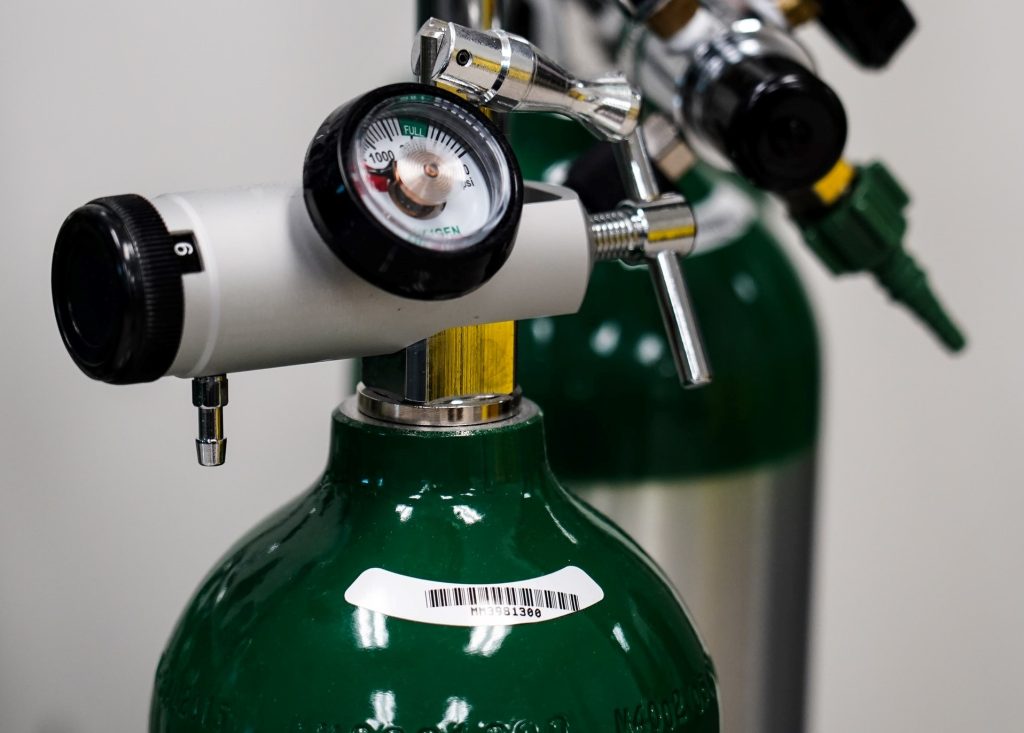
Supplies of medical oxygen in Ukraine are dangerously low due to disruption caused by the Russian invasion, the World Health Organization has warned.

Due to the crisis, the WHO estimates that the country needs an additional 20–25% increase in oxygen supplies over and above its normal needs. As it currently stands, the transport of oxygen cylinders across the country is being disrupted, especially into the capital Kyiv. As of 27 February, many hospitals across the country, including in Kyiv, had less than 24 hours’ supply remaining.

Furthermore, oxygen production facilities are experiencing shortages of zeolite, which is needed for the safe production of oxygen in the pressure swing absorption process

Prior to the conflict, the WHO had worked with Ukraine to improve its oxygen supply infrastructure, especially during the COVID pandemic. “Of the over 600 health facilities nationwide assessed by WHO during the pandemic, close to half were directly supported with supplies, technical know-how and infrastructure investments, enabling health authorities to save tens of thousands of lives,” the WHO said. This progress is threatening to be undone.

“Compounding the risk to patients, critical hospital services are also being jeopardised by electricity and power shortages, and ambulances transporting patients are in danger of getting caught in the crossfire,” the WHO said in its press release.

To offset this, the WHO is working through regional networks to bring in oxygen, as well as providing trauma treatment supplies. These would be brought in through a safe logistics corridor in Poland.

Médecins Sans Frontières (MSF) has announced that it is suspending activities in Ukraine. “These included care for people living with HIV in Severodonetsk; care for patients with tuberculosis in Zhytomyr; and improving access to healthcare access in Donetsk, in eastern Ukraine, where we have been providing much-needed healthcare, including for mental health, to conflict-affected communities,” the organisation said in an announcement.

However, it is working to ensure some continuity of its operations, and are working to provide trauma training to certain hospitals and have provided some trauma supplies.

The Ukrainian capital of Kyiv has also put out a call for donations of medicines, such as the antiviral amixin, the antibiotic nifuroxazide and the haemostatic agent aminocaproic acid.

Source: World Health Organization

To Properly Use AI to Analyse Breast Cancers, Look to Past Mistakes

Source: National Cancer Institute

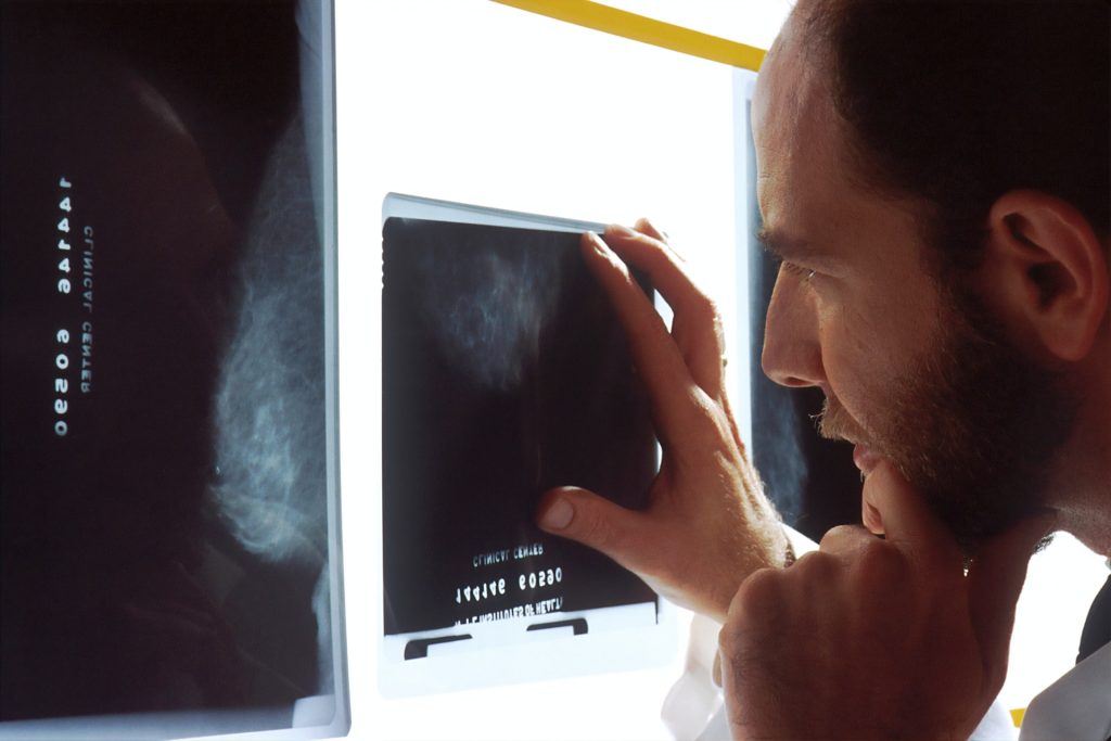
Doctors writing in an editorial in JAMA Health Forum caution that while using AI to analyse breast cancer tumours has the potential to improve healthcare efficiency and outcomes, similar technological leaps have previously led to higher rates of false-positive tests and over-treatment.

The editorial wasco-written by Joann G. Elmore, MD, MPH, professor of medicine at the David Geffen School of Medicine at UCLA, and Christoph I. Lee, MD, MS, MBA, a professor of radiology at the University of Washington School of Medicine.

“Without a more robust approach to the evaluation and implementation of AI, given the unabated adoption of emergent technology in clinical practice, we are failing to learn from our past mistakes in mammography,” the authors wrote.

One of those “past mistakes in mammography,” the authors said, was adjunct computer-aided detection (CAD) tools, which grew rapidly in popularity in the field of breast cancer screening starting more than two decades ago. CAD was approved by the FDA in 1998, and by 2016 more than 92% of U.S. imaging facilities were using the technology to interpret mammograms and hunt for tumours. However, CAD did not improve mammography accuracy., according to the evidence. “CAD tools are associated with increased false positive rates, leading to overdiagnosis of ductal carcinoma in situ and unnecessary diagnostic testing,” the authors wrote. The US Medicare system stopped paying for CAD in 2018, but by then the tools had run up more than $400 million a year in wasted health costs.

“The premature adoption of CAD is a premonitory symptom of the wholehearted embrace of emergent technologies prior to fully understanding their impact on patient outcomes,” Drs Elmore and Lee wrote. “As AI algorithms are increasingly receiving FDA clearance and becoming commercially available with ROC curves similar to what we observed prior to CAD clearance and adoption, how can we prevent history from repeating itself?”

The doctors suggest a number of safeguards to avoid “repeating past mistakes” such as tying reimbursement to proven efficacy.

Source: UCLA Health

Nintedanib Slows Autoimmune-related Lung Disease

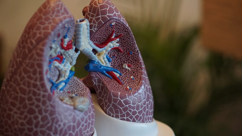
Anatomical model of lungs
Photo by Robina Weermeijer on Unsplash

Findings from a new clinical trial published in Arthritis & Rheumatology reveal that nintedanib,  an intracellular inhibitor of tyrosine kinases, may help patients with fibrosing from autoimmune disease-related interstitial lung diseases (ILDs).

ILDs are a common manifestation of systemic autoimmune diseases such as rheumatoid arthritis. Connective tissue diseases and vasculitides affect all areas of the lungs (bronchioles, parenchyma, alveoles) which is why ILD is a common feature of rheumatology diseases.

The trial enrolled 170 subjects with a fibrosing ILD other than idiopathic pulmonary fibrosis, with diffuse fibrosing lung disease of > 10% extent on high-resolution CT imagery, forced vital capacity (FVC) ≥ 45% predicted and diffusing capacity of the lungs for carbon monoxide ≥ 30% –< 80% predicted. FVC is a predictor of mortality in patients with autoimmune disease-associated ILDs.

Patients were randomised to receive nintedanib or placebo. Investigators assessed patients’ forced vital capacity (FVC). The trial found that the rate of decline in FVC over one year was -75.9 mL/year with nintedanib versus -178.6 mL/year with placebo.

“Until now, therapies that can significantly reduce the rate of decline in lung function in connective tissue disease–related ILDs characterised by progressive fibrosis have been lacking. We now have a therapeutic approach that offers a strategy for reducing the morbidity associated with these diseases,” said lead author Eric L. Matteson, MD, MPH, of the Mayo Clinic College of Medicine and Science.

Source: Wiley

A Synthetic Alternative to Pig-derived Heparin

Photo by Corinna Widmer from Pexels

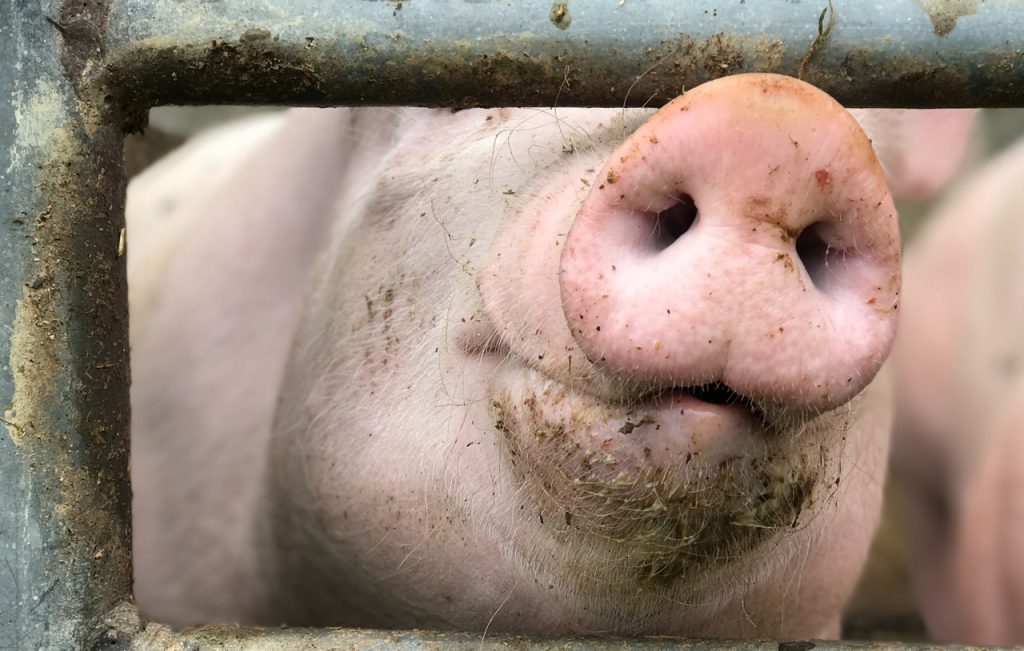
Scientists have developed a process to synthesise the vital blood thinner heparin, which is normally harvested from pig intestines. This synthetic heparin would help the quality control issues and shortages associated with pig-derived heparin.

The most expensive part of a pig is not a cut of bacon or a chop, but the part of the intestine used to make heparin. About 2000 pigs required to produce a kilogram of heparin, which provides medication to up to 6000 patients. In total, it is estimated that about one billion primarily Chinese food pigs each year also supply intestines for the extraction and processing of heparin.

However, this can cause problems for patients. When making medicines derived from animals, the chemical structure is rarely uniform. There are relatively common but harmless side effects, and in very rare cases severe and life-threatening immune reactions. Additionally, there are ethical and religious concerns for many patients. Bovine- and sheep-derived heparin are also produced but have the same concerns of being of animal origin. In fact, prior to 2000, heparin was derived from cows until the outbreak of mad cow disease.

Therefore, it has long been an ambition among researchers to make heparin in a laboratory to get cleaner heparin without side effects. Now researchers from the Copenhagen Center for Glycomics at the University of Copenhagen are ready with a study that shows that it is possible to make heparin without the use of animals.  which was published in Science Advances.

“By making heparin without the animal, you get a much cleaner and more uniform chemical structure. We show that we can do it in the laboratory, i.e. in a so-called ‘cell factory’, in the same way as many other types of medicine are made. It is a step in the direction of the development that has also happened with insulin, which was previously extracted from the pancreas in pigs before we learned to produce it in the laboratory,” explained Associate Professor Rebecca Miller, who led the study. 

There is already a synthetic alternative to heparin, but it is difficult to dose and can lead to overdose. Because of this, GPs often prescribe pig-derived heparin to their patients.

Heparin is today extracted from pig intestines’ mucosa. Due to the sheer number of patients who need the medicine, the scale of the production is vast, making quality control a recurring problem for manufacturers.

In 2008, a number of stocks of heparin from Chinese pigs were recalled when it was found that the medicine was contaminated. The case ended up costing the lives of more than 100 Americans.

“Of course you want to avoid that, in addition to moving the source from animals to laboratory cells. With our new technology, we have made a design for how to make heparin in a cell that is clean and uniform and it suggests that it has the same medicinal effect as market heparin. In this way, you potentially get a product that leads to neither common nor life-threatening side effects,” said Richard Karlsson, PhD, who has also contributed to the main author of the study.

Right now, the world is facing a shortage of heparin because swine flu has thinned the pig population in China, the largest heparin producer.
Next steps would be to scale up production to provide much larger quantities of the new synthetic heparin. 

Source: University of Copenhagen