Day: January 27, 2022

Scientists Succeed in Regrowing Frog Legs

Photo by Nature-for-Future from Pexels

Using a mix of drugs and a regenerative seal, scientists were able to successfully regrow frog legs, as reported in Science Advances. This represents an eventual step towards possibly regrowing limbs in humans.

On adult frogs, which are naturally unable to regenerate limbs, the researchers were able to trigger regrowth of a lost leg using a five-drug cocktail applied in a silicone wearable bioreactor dome that seals in the treatment over the stump for just 24 hours. That brief treatment sets in motion an 18-month period of regrowth that restores a functional, near-complete leg.

In humans and mammals loss of a large and structurally complex limb cannot be restored by any natural process of regeneration in humans or mammals. In fact, we tend to cover major injuries with an amorphous mass of scar tissue, protecting it from further blood loss and infection and preventing further growth.

The Tufts University researchers triggered the regenerative process in African clawed frogs by enclosing the wound in a silicone cap, which they call a BioDome, containing a silk protein gel loaded with the five-drug cocktail.

Each drug fulfilled a different purpose, including tamping down inflammation, inhibiting collagen production which would lead to scarring, and encouraging the new growth of nerve fibres, blood vessels, and muscle. The combination and the bioreactor provided a local environment and signals that tipped the scales away from the natural tendency to close off the stump, and toward the regenerative process.

A dramatic growth of tissue was observed in many of the treated frogs, re-creating an almost fully functional leg which was able to respond to stimuli, though the “toes” grown had no bones.

“It’s exciting to see that the drugs we selected were helping to create an almost complete limb,” said Nirosha Murugan, research affiliate at the Allen Discovery Center at Tufts and first author of the paper. “The fact that it required only a brief exposure to the drugs to set in motion a months-long regeneration process suggests that frogs and perhaps other animals may have dormant regenerative capabilities that can be triggered into action.”

Within the first few days after treatment, they detected the activation of known molecular pathways that are normally used in a developing embryo. Activation of these pathways could allow the burden of growth and organisation of tissue to be handled by the limb itself, similar to that in an embryo, rather than require ongoing therapeutic intervention over the many months it takes to grow the limb.

Animals naturally capable of regeneration live mostly in an aquatic environment. The first stage of growth after loss of a limb is the formation of a blastema, a mass of stem cells at the end of the stump, which is used to gradually reconstruct the lost body part. The wound is rapidly covered by skin cells within the first 24 hours after the injury, protecting the reconstructing tissue underneath.

“Mammals and other regenerating animals will usually have their injuries exposed to air or making contact with the ground, and they can take days to weeks to close up with scar tissue,” said Tufts University Professor David Kaplan, co-author of the study. “Using the BioDome cap in the first 24 hours helps mimic an amniotic-like environment which, along with the right drugs, allows the rebuilding process to proceed without the interference of scar tissue.”

Previous work using just progesterone with the BioDome resulted in a spike-like limb.

The five-drug cocktail is a major step toward the restoration of fully functional frog limbs and suggests further exploration of drug and growth factor combinations could lead to regrown limbs that are even more functionally complete.

“We’ll be testing how this treatment could apply to mammals next,” said corresponding author Professor Michael Levin at Tufts University.

“Covering the open wound with a liquid environment under the BioDome, with the right drug cocktail, could provide the necessary first signals to set the regenerative process in motion,” he said. “It’s a strategy focused on triggering dormant, inherent anatomical patterning programs, not micromanaging complex growth, since adult animals still have the information needed to make their body structures.”

Source: Tufts University

An Updated Look at the Link Between Alcohol and Epilepsy

Photo by Pavel Danilyuk on Pexels

A new meta-analysis has established an association between alcohol and epilepsy, in contrast to previous studies which reported conflicting results on the relationship.

Epilepsy is one of the most common neurological conditions, with an annual incidence of 40–70 per 100 000 people in industrialised countries. It is also a  disease that is highly stigmatised.

A number of studies have focused on how alcohol consumption leads to provoked seizures, commonly resulting from alcohol withdrawal, or heavy intoxication. Very few of these however focused on the link between alcohol consumption and unprovoked seizures. A 2010 meta-analysis found that alcohol users were more prone to developing unprovoked seizures – but data from recent cohort studies contradict these findings. A 2018 meta-analysis suggested that the relationship may only hold true for heavy drinkers.

Now, using more accurate diagnostic methods and recent data, a team of scientists from Pusan National University, South Korea, conducted an updated meta-analysis to conclusively clarify the relationship between alcohol consumption and unprovoked seizures and epilepsy.

For this meta-analysis, appearing in Drug and Alcohol Dependence, the researchers included a total of eight studies, of which five were case-control studies and three were cohort studies. They analysed the data to assess the dose-response relationship between alcohol intake and epilepsy. The results suggested that overall, compared to non-drinkers, alcohol drinkers were at a significantly higher risk of developing epilepsy, which increased with alcohol intake. These findings are consistent with previous meta-analyses.

An important finding was that cohort studies did not show a positive association between alcohol intake and epilepsy. In fact, 2 out of 3 cohort studies suggested that alcohol intake reduces the risk of epilepsy.

More large cohort studies are needed to prove a causal relationship between alcohol drinking and epilepsy, as well as a threshold of onset, said second author Professor Yun Hak Kim.

Source: EurekAlert!

NICD Warns of Malaria Being Misdiagnosed as COVID

Mosquito
Photo by Егор Камелев on Unsplash

The National Institute for Communicable Diseases has warned that, as South Africa enters its peak malaria season, cases of malaria are being misdiagnosed as COVID. Both malaria and COVID have similar non-specific early symptoms such as fever, chills, headaches, fatigue and muscle pain. Undiagnosed and untreated malaria rapidly progresses to severe illness and can be fatal.

Speaking at a media briefing on Wednesday, principal NICD medical scientist Dr Jaishree Raman said that Gauteng has seen a slight increase of malaria cases recently. 

Dr Raman noted that COVID “has pulled resources from the malaria programmes, reducing active surveillance and case investigation, which is reducing the ability [to] classify cases accurately.”

However, the NICD does not know the exact source of the malaria. “Data cleaning and case classification is ongoing, so at the moment, we cannot say whether the uptick in cases is due to locally-acquired or imported malaria,” she said.

The NICD advises that any individual that prevents with fever or ‘flu-like illness, if they reside in a malaria-risk area in Limpopo, KwaZulu-Natal and Mpumalanga or have travelled to a malaria-risk area, especially Mozambique, in the past six weeks, must be tested for malaria by blood smear microscopy or malaria rapid diagnostic test. If they test positive for malaria, the patient must be started on malaria treatment, immediately.

The NICD also advises patients to remember to inform their healthcare provider of their recent travel, especially to neighbouring countries and malaria risk areas in South Africa.  

‘Taxi malaria’, transmitted by hitch-hiking mosquitoes, should be considered in a patient with unexplained fever who has not travelled to a malaria-endemic area, but is getting progressively sicker, with a low platelet count.

Source: NICD

New Guidelines for Brain Cancer Care

Credit: National Cancer Institute

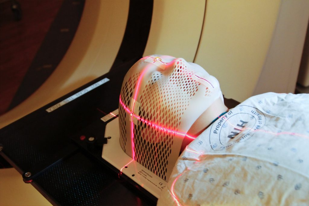
New guidelines for managing and treating brain metastases have been published in the Journal of Clinical Oncology and are set to improve care for cancer patients and extend and improve the quality of their lives.

The new guidelines come from an expert panel assembled by the American Society of Clinical Oncology (ASCO). The panel included a diverse range of top cancer doctors, as well as a patient representative.

The guidelines reflect the enormous advances in care for brain metastases  over the last few decades. In the 1970s, early attempts to develop guidelines largely emphasised steroids and whole-brain radiation therapy, without controlled, randomised studies to guide the use of surgery and chemotherapy.

Far more encompassing and far more evidence-based, the new guidelines will help doctors and patients make the best treatment decisions and achieve the best outcomes.

“When I started in this field 30 years ago, the average survival with brain metastases was 4 months, and most patients died from the brain disease. With improvements in therapies, fewer than one-quarter of patients die from the brain metastases, and some patients live years or are even cured,” said David Schiff, MD, a co-chair of the ASCO panel and the co-director of UVA Cancer Center’s Neuro-Oncology Center. “Equally importantly, the use of advanced localised radiation techniques and new targeted chemotherapies and immunotherapies have improved the quality of survival for most patients suffering from brain metastases.”

Up to 30% of cancer patients will have it spread to the brain, where it can be extremely difficult to treat. In the United States, approximately 200 000 new brain metastases are diagnosed each year.

The likelihood of a solid tumour spreading to the brain varies by cancer type, with approximately 20% of lung cancers spreading to the brain within a year after diagnosis. For patients with breast cancer, renal cell cancer or melanoma, that number is up to 7%. That is in addition to the patients found to have brain metastases at the time of their initial diagnosis.

Bringing together a diverse range of disciplines, the ASCO panel incorporated the results of more than 30 randomised trials published since 2008. The resulting guidelines cover everything from when surgery is appropriate and when and in what form radiation should be used to those circumstances in which medication alone may be employed.

The guidelines emphasise the importance of local therapies (surgery or stereotactic radiosurgery) for symptomatic brain metastases and lay out when these options are feasible. They highlight situations in which local therapy or whole brain radiotherapy can be deferred in place of chemotherapy, targeted therapy or immunotherapy depending on tumour type and molecular features. They also lay out how, in many cases, doctors can avoid the cognitive toxicity of whole brain radiotherapy by using either stereotactic radiosurgery or hippocampal-avoidant whole brain radiotherapy with the drug memantine.

“Patients with brain metastases may initially see a neurosurgeon, radiation or medical oncologist. The rigorous analysis underpinning these guidelines will provide each subspecialist a comprehensive picture of the treatment options appropriate for a given patient,” Dr Schiff said. “The result will allow patients the optimal personalised approach to maximise long-term control of brain metastases with good functional outcome.”

 Additional information is available at the ASCO website.

Source: EurekAlert!