Stepping patterns become slower and more variable when a person is not comfortable with their environment, researchers have found.
The findings, published in PLoS One, shows that the perceived comfort of an environment, rather than it being natural or not, affects how people walk, with potential lessons for urban design.
Lead author Daria Burtan of Bristol’s School of Psychological Science said: “Measuring the changes of a person’s walking patterns through an environment allows us to understand their experienced comfort on a moment-to-moment basis.
“This is an important step toward being able to objectively quantify the impact of particular architectural designs on people’s wellbeing.”
Research has shown that spending time in green spaces such as parks helps improve attention spans, concentration and wellbeing, which can be shown by improvements in measured stepping patterns when walking in different environments.
Daria added: “As our cognitive faculties begin to decline in older age, the stepping patterns we make with our feet become slower and more variable, relative to when we are younger in the prime of our health. We found that the same thing happened when people walked toward images of urban and nature scenes they didn’t feel comfortable with – their stepping patterns became slower and more varied, relative to when they were looking at scenes they found comfortable and which they liked.
“Not only does this suggest that environments in which we feel comfortable and safe, place fewer processing demands on our brains; it demonstrates how measuring the real-time dynamics of our gait provides us with a powerful new tool for informing on the cognitive impacts of architecture and urban design.”
The researchers are now seeking to understand which psychological factors contribute to sensory discomfort.
A series of studies in recent months has found that, thanks to the mRNA vaccine and previous infection, some people mount an extraordinarily powerful immune response against SARS-CoV-2 which some scientists have referred to as ‘superhuman’.
Called ‘hybrid immunity’, their bodies produce very high levels of antibodies, with great flexibility: likely capable of fighting off the SARS-CoV-2 variants currently circulating but also likely effective against future variants.
“Overall, hybrid immunity to SARS-CoV-2 appears to be impressively potent,” Crotty wrote in commentary in Science published in June.
“One could reasonably predict that these people will be quite well protected against most and perhaps all of — the SARS-CoV-2 variants that we are likely to see in the foreseeable future,” says Paul Bieniasz, a virologist at Rockefeller University who helped lead several of the studies.
Bieniasz and his colleagues found antibodies in these individuals capable of strongly neutralising the six variants of concern tested, including Delta and Beta, as well as several other viruses related to SARS-CoV-2, including SARS-CoV-1.
“This is being a bit more speculative, but I would also suspect that they would have some degree of protection against the SARS-like viruses that have yet to infect humans,” Bieniasz said.
People who have had a ‘hybrid’ exposure to the virus, were infected with it in 2020 and then immunised with mRNA vaccines this year. “Those people have amazing responses to the vaccine,” said virologist Theodora Hatziioannou at Rockefeller University, who also helped lead several of the studies. “I think they are in the best position to fight the virus. The antibodies in these people’s blood can even neutralize SARS-CoV-1, the first coronavirus, which emerged 20 years ago. That virus is very, very different from SARS-CoV-2.”
These antibodies were so effective they were even able to deactivate a virus purposefully engineered to be highly resistant to neutralisation, containing 20 mutations that are known to prevent SARS-CoV-2 antibodies from binding to it. Antibodies from those who were only vaccinated or who only had prior coronavirus infections were ineffecgtive against this engineered virus..
This shows how powerful the mRNA vaccine can be in those infected with SARS-CoV-2, she said. “There’s a lot of research now focused on finding a pan-coronavirus vaccine that would protect against all future variants. Our findings tell you that we already have it.
The catch is getting COVID. “After natural infections, the antibodies seem to evolve and become not only more potent but also broader. They become more resistant to mutations within the [virus].”
Hatziioannou and colleagues don’t know if this applies to all those mRNA-vaccinated and previously COVID-infected. “We’ve only studied the phenomena with a few patients because it’s extremely laborious and difficult research to do,” she said. “With every single one of the patients we studied, we saw the same thing.” The study reports data on 14 patients.
Several other studies lend credence to her hypothesis and reinforce the idea that exposure to both a coronavirus and an mRNA vaccine triggers an exceptionally powerful immune response. In one study in NEJM, scientists analysed antibodies generated by people who had been infected with SARS-CoV-1 back in 2002 or 2003 and who then received an mRNA vaccine this year.
Remarkably, these people also produced high levels of antibodies that could neutralise a whole range of variants and SARS-like viruses. Many questions remain, such as the effect of a third booster shot, or being infected again.
“I’m pretty certain that a third shot will help a person’s antibodies evolve even further, and perhaps they will acquire some breadth [or flexibility], but whether they will ever manage to get the breadth that you see following natural infection, that’s unclear.”
Immunologist John Wherry, at the University of Pennsylvania, is a bit more hopeful. “In our research, we already see some of this antibody evolution happening in people who are just vaccinated,” he said, “although it probably happens faster in people who have been infected.”
In a recent study, Wherry and colleagues showed that, over time, uninfected people with only two doses of the vaccine begin to produce more flexible antibodies, so a third dose would give even more of an evolutionary boost to the antibodies, Wherry said. So a person will be better equipped to fight off whatever variant the virus puts out there next.
“Based on all these findings, it looks like the immune system is eventually going to have the edge over this virus,” said Bieniasz, of Rockefeller University. “And if we’re lucky, SARS-CoV-2 will eventually fall into that category of viruses that gives us only a mild cold.”
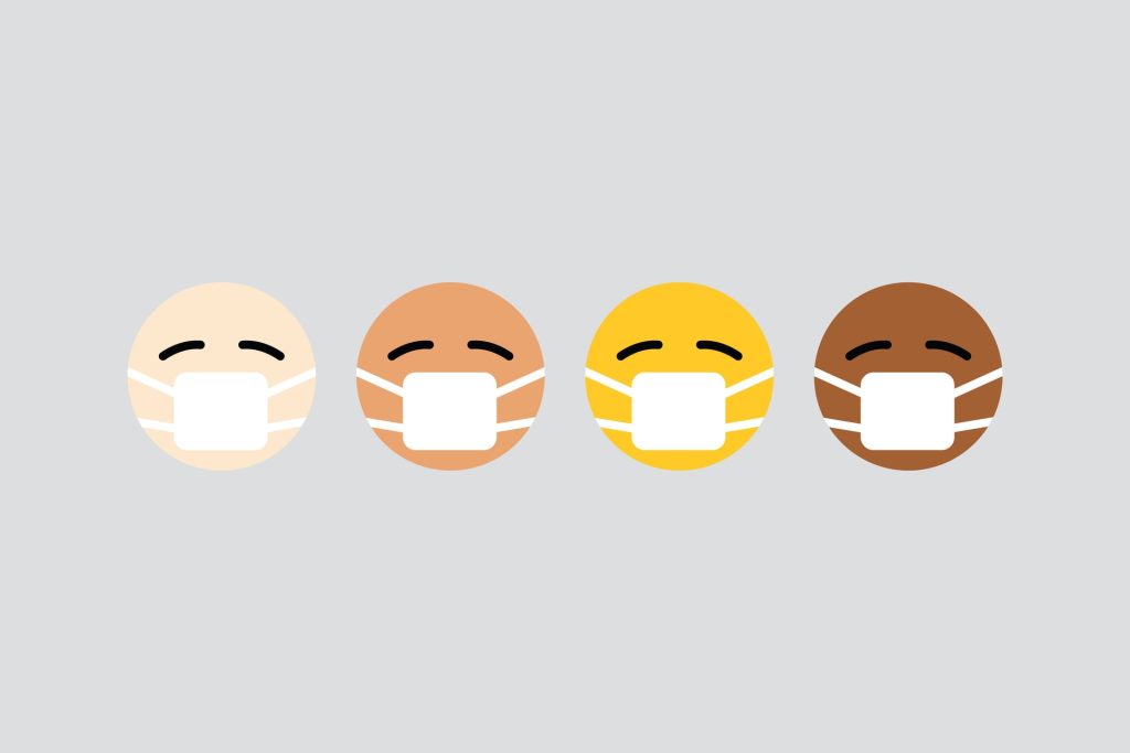
Emoji, those colourful symbols we use in WhatsApp and other communication applications, could be a valuable medical tool which lets patients better communicate symptoms, concerns, and other clinically relevant information, researchers argue.
In a commentary in the Journal of the American Medical Association, senior author Shuhan He, MD, an emergency department attending, advises that each medical discipline start to come up with its own unique set of iconography for official adoption and incorporation into everyday practice.
“The need to listen to patients is at the core of our mission as physicians, and the use of emoji is a great opportunity to take communication to another level,” said Dr He. “Emoji could be particularly important in treating children with still-developing language skills, people with disabilities that impair their ability to communicate, and the many patients who speak a different language.”
While around 3500 emoji are currently within the domain of the Unicode Consortium – the nonprofit organisation that maintains text standards across computers – only about 45 emoji can be considered relevant to medicine. The first, introduced in 2015, were the syringe and the pill. Apple added emoji in 2017 to represent people with disabilities, followed by symbols of the stethoscope, bone, tooth and microbe in 2019. He was co-creator of the anatomical heart and the lung emoji introduced globally in 2020 and is now working with colleagues, as well as with a wide range of medical societies and organisations to advocate for an additional 15 medically related emoji.
“It’s tempting to dismiss emoji as a millennial fad, but they possess the power of standardisation, universality and familiarity, and in the hands of physicians and other health care providers could represent a new and highly effective way to communicate pictorially with patients,” said Dr He. In emergency medical settings where time is critical, emoji could lead to a point-and-tap form of communication that could facilitate important clinical decisions, he adds. The tiny graphic symbols which now span all digital platforms – from mobile to tablet to desktop – could also have utility as annotations to hospital discharge instructions, which are often confusing if not incomprehensible to some patients.
The recent surge of telemedicine presents a great opportunity for medical emojis. It is well suited for patients visually conveying to healthcare providers the intensity of pain they have experienced over time, and for those providers to incorporate it into digital health records.
His research is on emoji to help patients and doctors communicate common symptoms – such as mobility, mood, and duration and quality of pain – that are associated with various diseases and conditions. “It’s clear that emoji have become part of the global, mainstream conversation, and that medical societies and physician committees and organisations need to take them seriously,” said Dr He. “Which means they should be determining now which emoji would best serve the interests of their patients, building consensus around the medical accuracy of these emoji, then working to get them approved through the global standard-setting body and working through the long adaptation and implementation process.”
A study into food purchasing behaviours shows that placing fruit and vegetables near store entrances and removing confectionery and other unhealthy products from checkouts and the end of nearby aisles prompts customers to make healthier food purchases.
The study, led by Dr Christina Vogel, Principal Research Fellow in Public Health Nutrition and Janis Baird, Professor of Public Health and Epidemiology at the University’s MRC Lifecourse Epidemiology Centre, was conducted in partnership with the national supermarket chain Iceland Foods Ltd. The trial took place in a number of Iceland stores in England, monitoring store sales as well as dietary patterns of sample customers.
The results showed confectionery sales decreased throughout the store while fruit and vegetable sales increased when non-food items and water were placed at checkouts and at the end of the opposite aisles, and an expanded fruit and vegetable section was repositioned near the store entrance. Beneficial effects were also observed for household fruit and vegetable purchasing and individual dietary quality. The findings are presented in the open-access journal PLOS Medicine.
Discussing the results of the study Dr Vogel said “Altering the layouts of supermarkets could help people make healthier food choices and shift population diet towards the government’s dietary recommendations. The findings of our study suggest that a healthier store layout could lead to nearly 10 000 extra portions of fruit and vegetables and approximately 1500 fewer portions of confectionery being sold on a weekly basis in each store.”
This research is more comprehensive than previous studies testing whether placement strategies can promote healthier food purchasing which have been limited in scope, for example including only a single location (ie at checkouts) or placing healthy and unhealthy products together. This study further aimed to reduce exposure of customers to calorie opportunities by placing non-food items at checkout and aisle-ends opposite and measuring effects on store sales, purchasing patterns on customer loyalty cards and the diets of more than one household member.
Matt Downes, Head of Format Development at Iceland, said: “We have been pleased to support this long-term study and the evaluation of how product placement in supermarkets can affect the diets of our customers. We know that childhood obesity is a growing issue and the retail industry has its part to play in tackling this. We hope that the outcomes of the study provide insights for the wider retail industry and policy makers about the impact of store merchandising on purchasing decisions.”
Prof Baird added “These results provide novel evidence to suggest that the intended UK government ban on prominent placement of unhealthy foods across retail outlets could be beneficial for population diet, and that effects may be further enhanced if requirements for a produce section near supermarket entrances were incorporated into the regulation.”
After 20 years of research, an established truth of how thrombin interacts with coagulation has been overturned.
“It has been said that an established truth in medicine lasts for about 10 years. It is probably the case that many truths last longer, but on the other hand, the time that different truths stand is constantly shrinking. This is because our perception of reality is changing rapidly, in step with new research,” said Tom Eirik Mollnes, Professor at the University of Oslo and Oslo University Hospital.
Based on more than 20 years of work to develop a whole blood model, Mollnes and colleagues have recently disproved an established truth about the immune system.
“There was an established truth in the literature for many years that a protein in the coagulation system called thrombin could activate a protein called C5 in the complement system,” Prof Mollnes said.
However, Prof Mollnes and his colleagues doubted whether the methods used in the studies were reliable.
“The notion that the protein thrombin could activate the protein C5 was only shown in so-called purified systems. That is, the proteins were taken out of their natural context,” he explained.
The researchers thought that the results would possibly be different if you looked at how the proteins work in their natural environment in the blood.
“The modified model made it possible to study the connection between the various proteins and defence systems as close to reality as possible,” Prof Mollnes explained. “Using the new model, we clearly showed that the previous findings were incorrect. We showed that the proteins changed structure and function during the purification, and that this was the reason for the former findings.”
When the proteins were in their natural environment in the blood, thrombin did not activate the protein in the complement system. Thus, the researchers had disproved the established truth.
“Many findings have been published in purified systems that are not representative of reality,” he said. “You can say that Gro Harlem Brundtland’s statement that “everything is connected to everything” is a very good description of how biology and the human body work. Therefore, it is important to use methods that make it possible to look at how different systems in the body interact and cooperate.”
The whole blood model makes such methods possible, and the model can be used widely. The whole blood model can, by and large, be used to study all the substances and biological systems in the blood.
Professor Mollnes therefore considers the model to have great potential.
“With the whole blood model, we have contributed to something that we will not only benefit from in our own laboratory, but that can be an asset to research groups in a number of fields,” he said.
It takes time to develop new models, and it was a long uphill battle for Prof Mollnes and his research group. Even so, the researchers have now received recognition for their work from the research community. The article got a recommendation by the editors of The Journal of Immunology, [PDF] in which it was published, as a ‘Top Reads Selection’.
“Changing so-called established truths is not easy, and we had to go through many rounds, with a number of experiments, to gain acceptance for our findings. That is why our work was especially recognised,” he concluded.
Mortality risk in sepsis is linked to the degree of platelet reduction, rather than absolute platelet count, according to new Japanese research.
Sepsis, a potentially life-threatening condition, arises from tissue and organ damage from an overactive infection response. Sepsis is commonly characterised by abnormally low platelet counts, which is believed to be associated with its high mortality rate.
Recently, Nagoya University researchers and colleagues have shown that a high degree of platelet reduction, rather than an abnormally low platelet count, raises mortality risks in sepsis. The findings, recently presented in the journal Scientific Reports, could lead to the development of precise and preventive treatments for sepsis-associated coagulopathy.
It is known that during sepsis, disseminated intravascular coagulation (DIC) forms tiny blood clots throughout the bloodstream, depleting platelets. Based on this, the international criterion for the diagnosis of sepsis-associated DIC uses platelet count and trials have been done using this criterion. However, very few trials have led to the development of effective treatments for sepsis-associated DIC.
There is however a different theory, that degree of platelet depletion (a rapid drop), rather than the absolute platelet count, accounts for much mortality risk in sepsis-associated DIC. But since there is little evidence for this theory, it has not been considered an international criterion for the disease prognosis.
With this in mind, researchers conducted a study to examine the significance of the degree of platelet reduction on sepsis mortality rate, using data from 200 859 sepsis patients staying in intensive care units of 208 US hospitals.
Corresponding author Dr Daisuke Kasugai of the Nagoya University Hospital, said: “To our knowledge, it was the largest study to evaluate the prognostic impact of both the degree of platelet depletion and absolute platelet counts in patients with sepsis.”
The degree of platelet reductions was found to be associated with the mortality risk associated with sepsis, regardless of absolute platelet count, indicating higher mortality risk with a fast decrease in platelet count. Dr Kasugai said: “Surprisingly, we also found that if the platelet count decreases by 11% or more, the risks of bleeding, as well as thrombosis development (a serious condition caused by the formation of blood clots in blood vessels or the heart), increases.”
The researchers therefore concluded that, compared to the absolute platelet count, the degree of platelet reduction could be a more plausible criterion for assessing the mortality risk of the sepsis-associated DIC. They hope that this study will lead to effective treatments for sepsis-associated DIC.
According to a new study, when rats are fed a high fat diet, this disturbs the body clock in their brain that normally controls satiety, leading to over-eating and obesity.
This new research, published in the Journal of Physiology, may be a cornerstone for future clinical studies that could restore the proper functioning of the body clock in the brain, to avoid overeating.
It was believed that the body clock resided only in the hypothalamus, but research over the years has clarified that some control of our body’s daily rhythms (hormone levels, appetite etc) lies in several other parts of the brain and body, including a group of neurons in the evolutionary ancient brainstem, called the dorsal vagal complex (DVC).
Specifically, the DVC has been shown to moderate food intake by inducing satiety. In obesity, research has shown that daily rhythms in food intake and the release of hormones related to eating, are blunted or eliminated. It is unclear if the malfunctioning of brain centres controlling appetite is a cause or the result of obesity.
This new study found that rats on a high-fat diet, before they started to gain weight, showed changes in the DVC’s daily neuronal rhythms and its response to appetite hormones. Thus, the researchers proposed that DVC disruption causes obesity.
Two groups of rats were used: those fed a well-balanced control diet (10% kcal from fat) and a high-fat diet (70% kcal from fat). To mimic the impact of unhealthy diet on humans, the researchers introduced the new diet to adolescent rats and monitored their food intake over 24h for four weeks.
Using multi-electrode arrays, the researchers measured DVC changes over 24h, simultaneously monitoring around a hundred DVC neurons from each brainstem slice. With this, circadian changes of neuronal activity could be assessed as well as neuronal responses to metabolically-relevant hormones in each of the diet groups.
Rats being nocturnal animals is a limitation of the study. The DVC activity peaked at the end of day, the rest phase for rodents, but an active phase for humans. Thus, it remains to be established if the phase of the brainstem clock is set to day and night, or whether it depends on patterns of rest and activity. These findings however could lead to understanding how to reset the body clock and tackle obesity.
First author Dr Lukasz Chrobok said:
“I’m really excited about this research because of the possibilities it opens up to tackle the growing health issue of obesity. We still do not know what are the time cues which are able to reset or synchronise the brainstem clock. Hopefully, the restoration of daily rhythms in this satiety centre before or after the onset of obesity may provide new therapeutic opportunities.”
This day, the first of its kind, is aimed at recognising and raising awareness of the invaluable role Field Epidemiologists play.
As health systems face increasingly complex threats, training workers in field epidemiology is even more important.. The NICD, a division of the National Health Laboratory Service, embarked on a joint collaboration more than 15-years ago in establishing the South African Field Epidemiology Training Programme (SAFETP). To date the program has trained 98 epidemiologists with the majority located in the public service in South Africa.
Field Epidemiologists, or ‘disease detectives’ are considered the cornerstone of public health preparedness and response. They undertake arduous, time-consuming tasks that include contact tracing, case investigations, community engagement, data collection and analysis.
One such ‘disease detective’ is Alain Musaka Abera, whose team was deployed to Equateur province in the DRC in response to an outbreak of Ebola virus disease (EVD). “The health zone of Ingende had already reported seven confirmed cases, including two deaths in the community,” said Abera, describing his work. “I had to set up the different pillars of epidemiological surveillance (management of alerts, active research, investigation, follow-up of contacts) and, at the same time, support coordination of the response in the health zone.
“The task was tough; the means of transport insufficient; communication almost non-existent. It was necessary to travel long distances in the forest on motorcycles that sometimes broke down and to cross the river in a canoe to search for and investigate suspects. It took courage, determination, and will to face these constraints.”
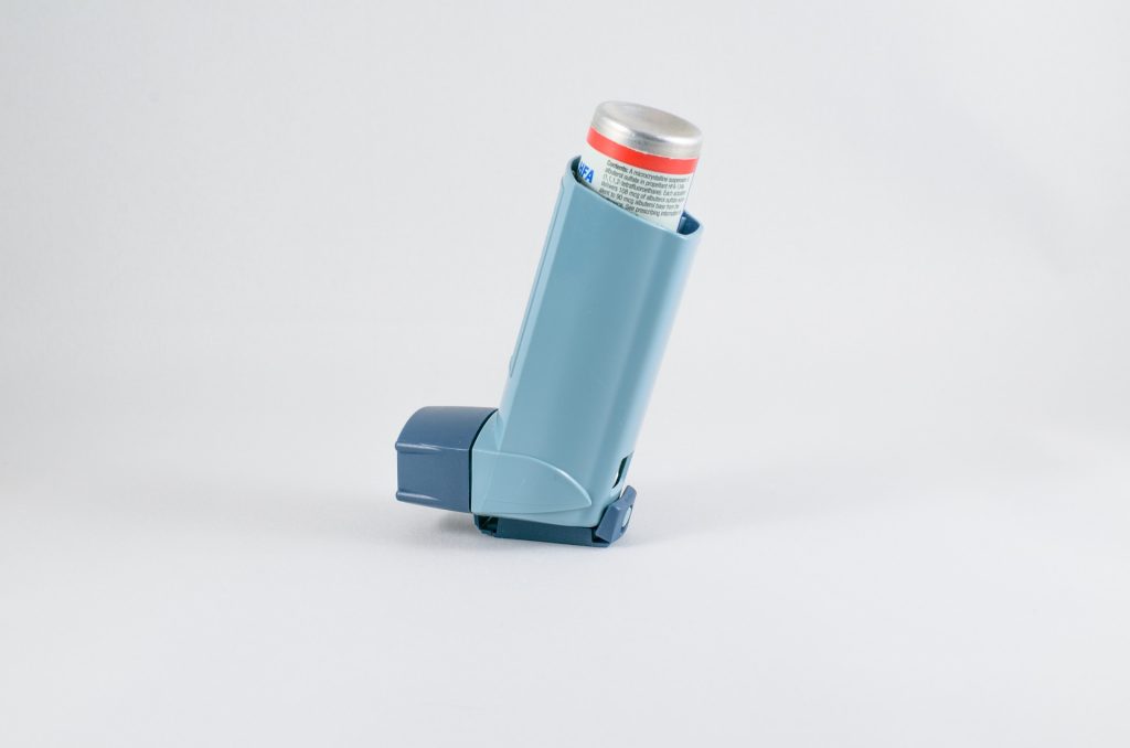
By pinning down the influence of the circadian system on nocturnal asthma, researchers have uncovered a key role for the biological clock in asthma.
Asthma severity has long been observed to worsen in the nighttime.Lung function is highest at around 4pm and worst around 4am. One longstanding question has been to what degree the body’s internal circadian clock contributes to worsening of asthma severity, as opposed to behaviours such as sleep. Using two circadian protocols, researchers have delineated the influence of the circadian system. Understanding the mechanisms behind asthma severity could have important implications for both studying and treating asthma.
“This is one of the first studies to carefully isolate the influence of the circadian system from the other factors that are behavioral and environmental, including sleep,” said co-corresponding author Frank AJL Scheer, PhD, director of the Medical Chronobiology Program in the Division of Sleep and Circadian Disorders at the Brigham.
As many as 75 percent of people with asthma report experiencing worsening asthma severity at night. Asthma severity is influenced by behavioural and environmental factors, such as exercise, air temperature, posture, and sleep environment. The researcher sought to understand the internal circadian system’s contributions to this problem. The circadian system is composed of a central pacemaker in the brain (the suprachiasmatic nucleus) and “clocks” throughout the body and is critical for the coordination of bodily functions and to anticipate the daily cycling environmental and behavioral demands.
To isolate the influence of the circadian system from that of sleep and other behavioural and environmental factors, the researchers enrolled 17 participants with asthma into two complementary laboratory protocols where lung function, asthma symptoms and bronchodilator use were continuously assessed. In the “constant routine” protocol, participants spent 38 hours continuously awake, in a constant posture, and under dim light conditions, with identical snacks every two hours. In the “forced desynchrony” protocol, participants were placed on a recurring 28-hour sleep/wake cycle for a week under dim light conditions, with all behaviours scheduled evenly across the cycle.
Co-corresponding author Steven A. Shea, Ph.D., professor and director at Oregon Institute of Occupational Health Sciences said, “We observed that those people who have the worst asthma in general are the ones who suffer from the greatest circadian-induced drops in pulmonary function at night, and also had the greatest changes induced by behaviours, including sleep. We also found that these results are clinically important because, when studied in the laboratory, symptom-driven bronchodilator inhaler use was as much as four times more often during the circadian night than during the day.”
A first-of-its-kind study on Alzheimer’s disease found an indication that omega-3 fatty acids taken early on protect against Alzheimer’s disease, despite not finding biomarkers in patients’ cerebrospinal fluid.
“We are careful not to draw any wider conclusions, but we can see a difference in the results of the memory tests. Patients who were taking omega-3 supplements at an early stage of the disease scored better,” cautioned Yvonne Freund-Levi, researcher in neuroscience at Örebro University.
The small study enrolled 33 patients, 18 of which were given omega-3 supplements morning and evening, and15 were in the control group. Spinal fluid samples were collected, and patients performed a memory test at the start of the study and after six months.
“We can see that the memory function of the patients in the group that had taken omega-3 is stable, whereas the patients in the control group have deteriorated. That’s what the memory tests show,” said Yvonne Freund-Levi.
“But we can’t see any differences between the groups when we look at the various biomarkers in the spinal fluid samples.”
However there are differences within the group given omega-3: an increase of two of the markers that are linked to damaged nerve cells. There is no clinical link to the memory tests, however.
“Even if this data isn’t enough for us to change our recommendations to patients at this time, it is an interesting material for researchers to build on.”
This study is based on a larger study with over 200 patients with mild to moderate Alzheimer’s disease, initiated by Yvonne Freund-Levi and her research team 15 years ago. In that previous study, the researchers found that omega-3 transfers from the supplements to the brain.
“We are cautious about giving recommendations, but we know that starting early is by far the best thing – it is difficult to influence the disease at a later stage. The best piece of advice we have to offer at the moment is to be physically active and to include omega-3 in your diet – in the form of oily fish or as supplements.”
In future, researchers will be able to measure biomarkers in blood samples rather than having to perform spinal tap procedures.
“We have already tested this approach at Sahlgrenska University Hospital. Without a doubt, it is so much better for the patients.”