Day: November 24, 2021

Understanding Mechanisms of Antibiotic Resistance

Source: NCI

If nothing is done, the problem of multidrug-resistant bacterial infections could be catastrophic by 2050, killing nearly 10 million people each year, according to experts’ predictions.

One person seeking solutions is Joseph Boll, assistant professor of biology at The University of Texas at Arlington, to identify and inhibit the defense mechanisms of Acinetobacter baumannii, a common pathogen in hospitals and clinical settings.

A. baumannii can cause infections in the blood, urinary tract and lungs, or in wounds in other parts of the body. Antibiotics are usually used to treat the infections, but many strains are resistant, including drugs of last resort, carbapenems.

“In previous research, we discovered that when A. baumannii experiences stress, such as antibiotic treatment, it modifies its cell envelope to tolerate the antibiotic for extended periods of time,” Prof Boll said. “Specific modifications allow the bacteria to survive long enough to acquire true antibiotic resistance, which can lead to antibiotic treatment failure. This can happen within 24 hours of antibiotic exposure.”

His team expects to identify what adaptations in the cell envelope allow the pathogen to survive in the presence of antibiotics and how survival contributes to the acquisition of true resistance.

In a recent study published in mBio, the team demonstrated that two LD-transpeptidase enzymes remodel A. baumannii’s cell envelope to promote its survival when under stress, such as the kind experienced during antibiotic treatment.

With this breakthrough, Hannah Bovermann, a senior double-major in biology and microbiology, is dissecting the genes that encode the bacteria’s LD-transpeptidases to learn what stress conditions induce their activation. She isolates the LD-transpeptidase promoters, the part of the DNA that controls when other parts of DNA are used, and glues it to a different gene whose function is to turn the bacterial cell blue. When the cell is in an environment where it wants to modify its cell envelope to protect itself, it turns blue, letting her observe the timing of the change.

To provoke this reaction, she administers antibiotics, experiments with various temperature changes, exposes the cell to pH gradients and subjects the cell to nutrient deprivations.

“Each response brings us closer to an understanding of how cell envelope modifications keep the bacterial cell intact in stress,” Bovermann said.

The researchers hope to find new targets on the cell surface for antibiotics to attack, strengthening existing medications’ potency against A. baumannii infections.

Clinicians have been pushed into using combinatorial therapies, where multiple drugs are employed to treat bacterial infections, but even those methods are becoming increasingly ineffective, Prof Boll said.

“It has become a game. Researchers discover a new antimicrobial, then bacteria become resistant to it. We are running out of options,” Prof Boll said. “Bacterial resistance is quickly outpacing new antibiotic development.”

Source: EurekAlert!

A Surprising Use for Ivermectin in Fighting West Nile Virus

Mosquito
Photo by Егор Камелев on Unsplash

Putting ivermectin in bird-feeders in places where Culex mosquitoes congregate showed promise in reducing the number of infectious mosquitoes that could transmit West Nile virus to humans, according to a researcher presenting at the American Society of Tropical Medicine & Hygiene (ASTMH) virtual meeting.

A pilot trial found that when ivermectin-treated bird-feeders were placed in contiguous lots, there was up to a 16% additional reduction in infectious mosquito days compared to when the bird-feeders were randomly placed, where there was only a 5% additional decline, reported Karen Holcomb, PhD, of the University of California Davis.

Furthermore, with just 33% coverage of ivermectin-treated bird-feeders in a neighbourhood, infectious mosquito days fell by 15% to 45%, she stated.
Since no human vaccine exists for West Nile Virus, the primary strategy has been vector control, she added. However, insecticide has a low specificity in targeting mosquitoes involved in West Nile transmission, and Holcomb also discussed some “non-targeted effects” of spraying in the human population.

Her group hypothesised that because ivermectin could kill mosquitoes while being minimally toxic in mammals and birds, it might be possible to treat bird-feeders with ivermectin, where the mosquitoes would bite the birds, ingest the ivermectin, and die before they could pass the virus on to humans.

Two earlier studies laid the groundwork: one that treated chickens with ivermectin and found a decrease in seroconversion and fewer older mosquitoes near the treated flocks, and an increase in mortality of mosquitoes following a blood meal on treated chickens.

Nevertheless, “the link between ivermectin and West Nile virus transmission was not fully elucidated,” Dr Holcomb said, adding there was no significant difference in mosquito abundance or infection prevalence, as well as variable serum concentrations in chickens.

Next steps of the study included determining what type of set-up worked best for ivermectin-treated bird-feeders in neighborhoods: either contiguous (with all treated bird-feeders in a row) or random. Dr Holcomb’s group found similar reductions in infections in mosquitoes and birds, but the greatest reductions in treated lots were from contiguous, not random placement, she said.

Uncertainty about ivermectin-induced mortality in wild mosquitoes remains, as well as the proper dose of ivermectin to induce mortality.

As a result of ivermectin’s controversial demand during the COVID pandemic, Dr Holcomb said she encountered logistical hurdles.

“During the past year, our collaborators noted it’s become harder to obtain ivermectin, and the ivermectin they were getting was lower quality than prior to COVID,” she told MedPage Today.

In any case, this strategy won’t be used in field control trials for at least a couple more years, Dr Holcomb noted, and “during that time, there should be a reduction in demand for ivermectin.”.

Source: MedPage Today

How Shigella Bacteria Hijacks Cells

Shigella bacteria Credit: S Bhimji, MD

Shigella, a bacterial pathogen that causes dysentery and is the leading cause of childhood diarrhoeal diseases, inserts a pore called a translocon into an infected person’s intestinal cells and then injects bacterial proteins into the cells. Once inside, the proteins hijack the cellular machinery to help Shigella multiply. In a study published in mBio, researchers report important details about Shigella’s translocon, which may help researchers develop an effective strategy to block this critical component of infection.

“Shigella infects our gut by manipulating our intestinal cells and tricking them into letting the Shigella inside. In fact, there are many bacterial pathogens that use this same, or similar, mechanism to infect us,” said lead author Poyin Chen, PhD, a postdoctoral fellow at MGH. “This translocon pore is essentially the gateway through which bacterial proteins get pumped into our cells. We know that this structure is made of two proteins – IpaB and IpaC – but what we don’t know is how these proteins fit together to make this pore.”

Using protein mapping techniques to look closely at translocons when they were embedded in cell membranes, the researchers were able to see that ipaB makes up the inner ring of the pore. “If you think of the translocon pore as a donut, this would be the walls of the donut hole. This finding is important because this is the part of the translocon pore that directly interacts with bacterial proteins as they are injected into our cells,” Dr Chen explained. “With the findings from this study, we can begin to understand if this pore acts as a slippery tube that bacterial proteins travel through or if the translocon pore can control the flow of bacterial proteins into our cells.”

Such details may help investigators target the translocon and block the entry of Shigella proteins into cells. “For something that is so essential to establishing infection, we know terribly little of how it’s made and how it works,” said Dr Chen. “As we gain a better understanding of its parts, we will be able to approach the structure as a whole and maybe even find ways to neutralise the function of this structure to prevent infection before it can begin.”

Source: Massachusetts General Hospital

South Africa Faces Vaccine Glut as Uptake Slows

Photo by Mat Napo on Unsplash

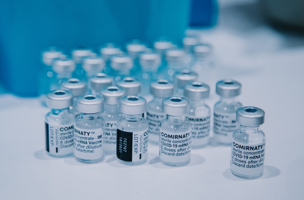
South Africa has asked Johnson & Johnson and Pfizer to delay delivery of COVID vaccines as it has too much stock now, health ministry officials said, as vaccine hesitancy continues to slow the immunisation campaign.

About 35% of South Africans are fully vaccinated, still only half the government’s target of 70% by year end. In the past 15 days, an average of 106 000 doses a day have been administered. At the beginning of the year, the programme had been beset by a lack of doses for a wide range of reasons, from AstraZeneca’s ineffectiveness against the Beta variant to overseas production delays. 

Deputy director-general of the Health Department, Nicholas Crisp, told Reuters that South Africa had 16.8 million doses in stock and said that deliveries had been deferred.

A spokesman for the Health Ministry said: “We have 158 days’ stock in the country at current use. We have deferred some deliveries.”

Stavros Nicolaou, chief executive of Aspen Pharmacare, which is packaging 25 million doses a month of J&J vaccines in South Africa, said most of the vaccines bound for South Africa would now be diverted to the rest of Africa, and deliveries would likely be deferred until the first quarter of next year.

A Pfizer spokesperson said: “We remain adaptable to individual country’s vaccine requirements whilst continuing to meet our quarterly commitments as per the South Africa supply agreement.”

The government has been trying to boost the rate of daily administered doses, such as with R100 ‘Vooma vouchers’ for registering to vaccinate, but even these have failed to sufficiently stoke uptake.

“There is a fair amount of apathy and hesitancy,” said Wits University’s Professor Shabir Madhi.

On Twitter, he further suggested using the excess stock for booster shots, which would “provide all single dose JJ adult recipients a JJ or Pfizer boost, and  those > 65 or immunosuppressive conditions an additional Pfizer dose if received 2 doses > 5 months ago.” 

Source: U.S. News

Significant White Matter Changes in Autism Revealed by MRI

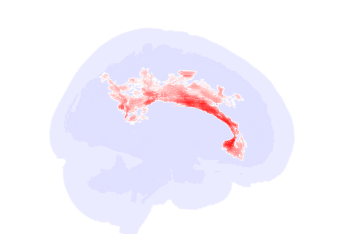
Significant alterations in the brain’s white matter in adolescents with autism spectrum disorder (ASD). Credit: RSNA and researcher, Clara Weber

Using specialised MRI, researchers found significant changes in the microstructure of the brain’s white matter, especially in the corpus callosum in adolescents and young adults with autism spectrum disorder (ASD) compared to controls. This research will be presented next week at the annual meeting of the Radiological Society of North America (RSNA).

“One in 68 children in the U.S. is affected by ASD, but high variety in symptom manifestation and severity make it hard to recognise the condition early and monitor treatment response,” explained Clara Weber, postgraduate research fellow at Yale University School of Medicine. “We aim to find neuroimaging biomarkers that can potentially facilitate diagnosis and therapy planning.”

Researchers reviewed diffusion tensor imaging (DTI) brain scans from a large dataset of patients between the age of six months and 50 years. DTI is an MRI technique that measures connectivity in the brain by detecting how water moves along its white matter tracts. Water molecules diffuse differently through the brain, depending on the integrity, architecture and presence of barriers in tissue.

“If you think of gray matter as the computer, white matter is like the cables,” Weber said. “DTI helps us assess how connected and intact those cables are.”

For the study, clinical and DTI data from 583 patients from four existing studies of distinct patient populations were analysed: infants (median age 7 months), toddlers (median age 32 months), adolescents, and young adults.

“One of the strengths of our study is that we looked at a wide range of age groups, not just school-aged children,” Weber said.

To assess the influences of age and ASD diagnosis on white matter microstructure, the research team created fractional anisotropy, mean diffusivity and radial diffusivity maps using data from the four studies.

Fractional anisotropy is the extent water diffusion is restricted to just one direction. A value of zero means that diffusion is unrestricted in all directions, while one means that diffusion is unidirectional. Mean diffusivity is the overall mobility of water molecules, indicating how densely cells are packed together. Radial diffusivity is the extent water diffuses perpendicular to a white matter tract.

“When white matter integrity is disrupted, we see more water diffusing perpendicularly, which translates to a higher radial diffusivity,” Weber said.

The key finding of the analysis was reduced fractional anisotropy within the anterior/middle tracts of the corpus callosum in adolescent and young adult ASD patients compared to individuals in the control group. The corpus callosum is a thick bundle of nerve fibers that connects and allows the two sides of the brain to communicate. Corresponding increases in ASD-related mean diffusivity and radial diffusivity were found in young adults.

“In adolescents, we saw a significant influence of autism,” Weber said. “In adults, the effect was even more pronounced. Our results support the idea of impaired brain connectivity in autism, especially in tracts that connect both hemispheres.”

Compared to controls, no reduction in fractional anisotropy was seen in the same tracts in toddlers and infants with ASD.

The researchers hope the findings can help improve early diagnosis of ASD and provide potential objective biomarkers to monitor treatment response.

“We need to find more objective biomarkers for the disorder that can be applied in clinical practice,” Weber said.

Source: EurekAlert!

IBS Dietary Therapy Works Best with Certain Gut Microbiomes

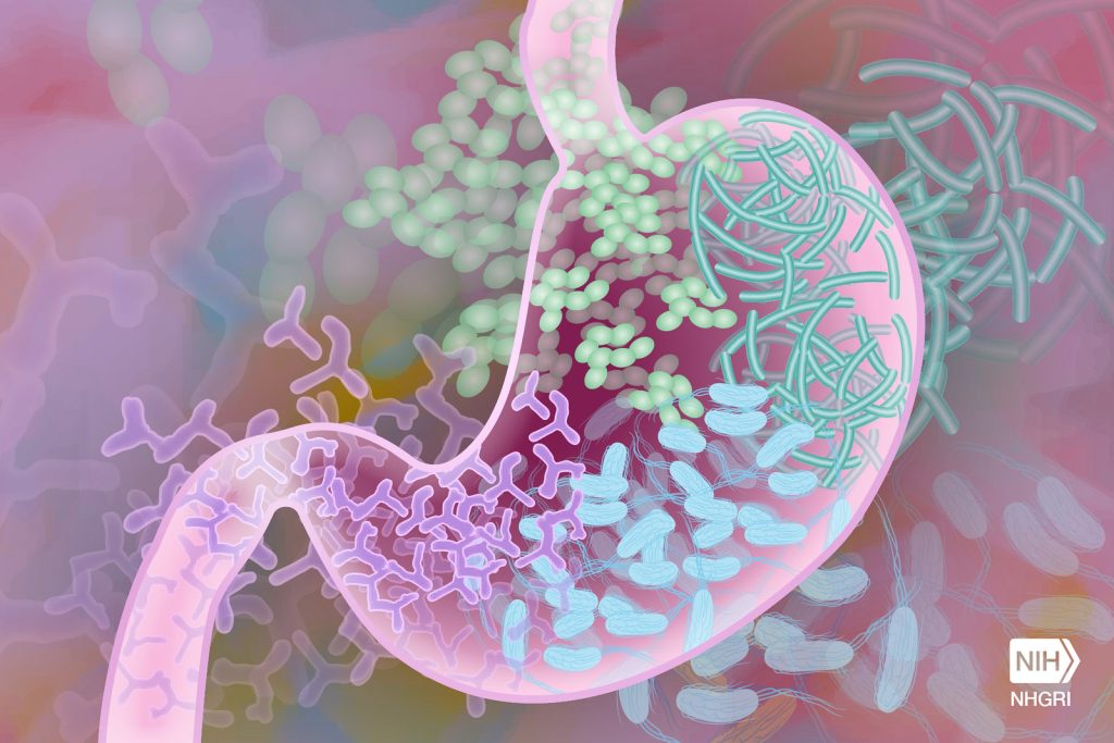
Beneficial gut bacteria. Credit: Darryl Leja, National Human Genome Research Institute, National Institutes of Health

People who respond well to the irritable bowel syndrome (IBS) dietary therapy of reduced fermentable carbs have an abundance of particular types of bacteria in their gut, reveals research published online in the journal Gut.

The composition of the gut microbiome is thought to have a major role in the development of IBS. Restricting fermentable carbs, found in many foods including wheat, onions, and milk, is usually recommended to ease symptoms, an approach known as the low FODMAP (fermentable oligo-, di-, mono-saccharides and polyols) diet. But why this diet works is not fully understood.

In a bid to fill this knowledge gap, the researchers analysed stool samples of 56 people with IBS and 56 people who lived with them, but without the condition, to identify the microbial profile and genes involved in converting food into active molecules while on their usual diet.

They then assessed the clinical response in 41 of these pairs after 4 weeks on the low FODMAP diet by reviewing their stool samples again.

Before adoption of the low FODMAP diet, analysis of the stool samples of those with IBS revealed two distinct microbial ‘signatures’, which the researchers referred to as ‘pathogenic-like’ (IBSP) and as ‘health-like’ (IBSH).

The pathogenic microbial signature was abundant in harmful Firmicutes sp, including known disease causing bacteria, such as C. difficileC. sordellii and C. perfringens, but very low in beneficial Bacteroidetes species.

The lactic acid bacteria Streptococcus parasanguinis and Streptococcus timonensis that are usually found in the mouth were also abundant. And bacterial genes for amino acid and carbohydrate metabolism were overexpressed, which may explain the excess of some metabolites that are linked to IBS symptoms, say the researchers.

The healthy microbial signature of the other IBS patients was similar to that found in the comparison group (household members).

After 4 weeks on the low FODMAP diet, the microbiome of the comparison group and those with the healthy microbial profile stayed the same.

But the microbiome of those with the pathogenic profile became healthier, with an increase in Bacteroidetes, and a fall in Firmicutes species. And the bacterial genes involved in the metabolism of amino acids and carbs were no longer overexpressed.

In 3 out of 4 of IBS patients, symptoms improved. But the clinical response to the low FODMAP diet was greater in those with IBS and a pathogenic microbial signature than it was in those with IBS and a healthy microbial signature in their gut.

“The evidence associating diet, the microbiome and symptoms in [pathogenic IBS] is compelling, but studies following the introduction of candidate organisms into an animal model are needed to prove the relationship is causal,”  the researchers cautioned.

Nevertheless, they suggest their findings could lead to a microbial signature to identify those who would respond best to a low FODMAP diet and better manage those who wouldn’t.

“If the bacteria represented in the [pathogenic] subtype are shown to play a pathogenic role in IBS, perhaps through their metabolic activity, this provides a target for new therapies and an intermediate [marker] by which to assess them,” they suggest.

In a linked editorial, Professor Peter Gibson and Dr Emma Halmos of Melbourne’s Monash University, describe the introduction and adoption of the FODMAP diet as “a major change in the management of patients with irritable bowel syndrome (IBS) towards integrated care.

But while “an effective symptomatic therapy, [it’s] one that carries risks associated with exacerbating disordered eating, challenging nutritional adequacy and putatively inducing dysbiotic gut microbiota,” they added.

They point out some limitations of the research, including that FODMAP intake was poorly assessed, fibre intake,which can also influence the microbiome wasn’t reported and patient drop-out reduced the power of the study.

Nevertheless, the authors concluded that “the beauty of [the study] is not in its definitive nature, but that it enables the creation of feasible innovative hypotheses that can be examined by focused studies. Perhaps the FODMAP diet is not just a symptomatic therapy.” 

Source: BMJ

The Search for New Cancer Therapies Strikes Gold

Photo by Jingming Pan on Unsplash

The gold complex auranofin has traditionally been used for treating rheumatism but is also being evaluated as a one number of new cancer therapies. According to a study published in Redox Biology, molecules with the same inhibition effect have been discovered that have a more specific effect than auranofin and therefore may have greater potential as cancer therapies.

Auranofin (AF) is classed by the WHO (World Health Organization) as an anti-rheumatic agent and is an active component in the drug Ridaura. AF is also currently being assayed in a string of clinical trials as a possible cancer therapy. One reason for the researchers’ interest in AF is its ability to inhibit thioredoxin reductase (TrxR), a protein central to the thioredoxin system, which protects cells from oxidative stress in all mammals. 

However, TrxR also protects cancer cells, making cancer therapies less effective. Moreover, TrxR, which affects cellular growth and survival, is upregulated in certain forms of cancer.

“There’s a great deal of interest in the ability to inhibit the thioredoxin system in the treatment of cancer, but there’s a risk that healthy cells will also be damaged and killed,” says the study’s co-last author Elias Arnér, professor at the Department of Medical Biochemistry and Biophysics at Karolinska Institutet. “Our aim is for TrxR inhibitors to be as specific as possible.”

The researchers studied the effects of AF in mouse cancer cells (lung adenocarcinoma and melanoma) and compared them with other recently-developed TrxR-inhibiting molecules called TRi-1 and TRi-2 (thioredoxin reductase inhibitors 1 and 2). 

The study, which was based on new proteomic methods of analysing the entire set of proteins in cells, suggests that the TRi compounds are more specific in their effect than AF. The results show that AF causes very high levels of oxidative stress and has other effects that seem unrelated to the inhibition of TrxR. They also demonstrate that TRi-1 seems to be the most specific TrxR inhibitor so far.

“Our results can serve as an important blueprint for further studies of AF’s mechanism of action and side effects,” said the study’s other co-last author Roman Zubarev, professor at the Department of Medical Biochemistry and Biophysics, Karolinska Institutet. “Having now compared AF with the more specific molecules TRi-1 and TRi-2, we hope that our findings will contribute to the further development of TrxR inhibitors as anticancer drugs.”

Source: Karolinska Insitutet

Immunity to Other Coronaviruses Confers COVID Protection

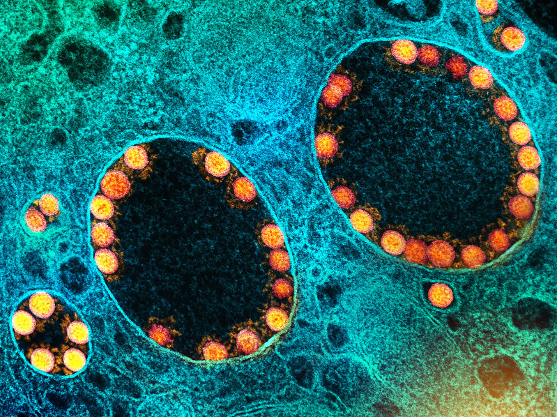
Transmission electron micrograph of SARS-CoV-2 virus particles (gold) within endosomes of a heavily infected nasal Olfactory Epithelial Cell. Image captured at the NIAID Integrated Research Facility (IRF) in Fort Detrick, Maryland. Credit: NIAID

Researchers have discovered another component beside previous infection or vaccination that contributes to SARS-CoV-2 immunity – previous antibody responses to other, harmless coronaviruses. “People who have had strong immune responses to other human coronaviruses also have some protection against SARS-CoV-2 infection,” said Alexandra Trkola, head of the Institute of Medical Virology at University of Zurich.

The study, published in Nature Communication, used a specially developed assay to analyse antibody levels against four other human coronaviruses in 825 serum samples taken before  the emergence of SARS-CoV-2, as well as 389 samples from donors infected with the virus. Combining these analyses with computer-based models enabled the team to precisely predict how well the antibodies would bind to and neutralise invading viruses.

The researchers were able to demonstrate that people who caught SARS-CoV-2 had lower levels of antibodies against coronaviruses that cause common colds compared to uninfected people. In addition, people with high levels of antibodies against harmless coronaviruses were less likely to have been hospitalized after catching SARS-CoV-2. “Our study shows that a strong antibody response to human coronaviruses increases the level of antibodies against SARS-CoV-2. So someone who has gained immunity to harmless coronaviruses is therefore also better protected against severe SARS-CoV-2 infections,” says Trkola. This type of immune response is referred to as cross-reactivity, and it also occurs with T cell responses,  the additional line of the immune system in the defense against infections.

People are only fully protected against SARS-CoV-2 shortly after they have recovered from an infection or have received an effective vaccination. This is when antibody levels against the virus are still very high. As these levels drop over time, infection is no longer prevented, but the immunological memory quickly reactivates the body’s defenses, the production of antibodies as well as the T cell defense. “Of course, immune responses targeting SARS-CoV-2 that are mounted by the memory cells are far more effective than cross-reactive responses. But even though the protection isn’t absolute, cross-reactive immune responses shorten the infection and reduce its severity. And this is exactly what is also achieved through vaccination, just much, much more efficiently,” said Trkola.

Whether cross-reactivity also works in the opposite direction is not yet known. “If SARS-CoV-2 immunity also offers some degree of protection from infection with other coronaviruses, we would be a significant step closer to achieving comprehensive protection against other coronaviruses, including any new variants,” the virologist explains. This idea is also supported by the fact that cross-reactive protection is not only based on antibodies, but very likely also on T cells.

Source: University of Zurich

Heart Failure Risk Further Increased by Aspirin Use

Aspirin may increase heart failure risk in at-risk people
Photo by Stephen Foster on Unsplash

Aspirin use is associated with a 26% higher risk of heart failure in people with at least one risk factor for it, according to a study published today in ESC Heart FailureRisk factors included smoking, obesity, hypertension, high cholesterol, diabetes, and cardiovascular disease.

“This is the first study to report that among individuals with a least one risk factor for heart failure, those taking aspirin were more likely to subsequently develop the condition than those not using the medication,” said study author Dr. Blerim Mujaj of the University of Freiburg, Germany. “While the findings require confirmation, they do indicate that the potential link between aspirin and heart failure needs to be clarified.”

The influence of aspirin on heart failure is controversial, and so the study sought to investigate its association with heart failure incidence in people with and without heart disease and assess whether it is related to a new heart failure diagnosis in at-risk individuals.

The analysis included 30 827 individuals at risk for developing heart failure who were enrolled from Western Europe and the US into the HOMAGE study. The definition of “at risk” included one or more of the following: smoking, obesity, hypertension, high cholesterol, diabetes and cardiovascular disease. Participants were aged 40 years and older and were free of heart failure at baseline. Aspirin use was recorded at enrolment and participants were classified as users or non-users. Participants were followed-up for the first incidence of fatal or non-fatal heart failure requiring hospitalisation.

Average participant age was 67, 34% were women, and at baseline, a total of 7,698 participants (25%) were taking aspirin. During the 5.3-year follow-up, 1330 participants developed heart failure.

The investigators assessed the association between aspirin use and incident heart failure after adjusting for factors including demographic variables, medical history and medication. Taking aspirin was independently associated with a 26% raised risk of a new heart failure diagnosis.

For consistency, the researchers repeated the analysis after matching aspirin users and non-users for heart failure risk factors. In this matched analysis, aspirin was associated with a 26% raised risk of a new heart failure diagnosis. After excluding patients with a history of cardiovascular disease, in 22 690 participants (74%) without cardiovascular disease, aspirin use was still associated with a 27% increased risk of incident heart failure.

Dr Mujaj noted that “this was the first large study to investigate the relationship between aspirin use and incident heart failure in individuals with and without heart disease and at least one risk factor. Aspirin is commonly used – in our study one in four participants were taking the medication. In this population, aspirin use was associated with incident heart failure, independent of other risk factors.”

He concluded that “large multinational randomised trials in adults at risk for heart failure are needed to verify these results. Until then, our observations suggest that aspirin should be prescribed with caution in those with heart failure or with risk factors for the condition.”

Source: European Society of Cardiology