Month: October 2021

‘Sutrodes’ Could Treat Spleen Conditions Using Electricity

Photo by Zoltan Tasi on Unsplash

Using a flexible ‘sutrode’ – a combination of suture and electrode – a group of researchers has advanced a way to treat spleen conditions by applying only electrical current.

Electroceuticals, where electrical stimulation is used to modify biological functions, could minimally invasively treat medical conditions and result in few side effects.  

The work, which appears in the Nature Journal of Communications Biology, builds on previous studies when the team introduced the sutrode to the world just over a year ago. This graphene-based electrode is an electrical stimulation device that could replace the use of pharmaceuticals to treat a range of medical conditions. The sutrode, created using a technique called fibre wet spinning, has an electrode’s electrical properties and a suture’s mechanical properties.

“The flexibility and superb sensitivity of the sutrode is allowing us to expand our understanding of how the nervous system controls main body organs, a critical step towards developing advanced therapies in bioelectronic medicines,” reported the study leader, Professor Romero-Ortega. “Our collaborative work uncovered that the spleen is controlled by different terminal nerves, and that the sutrode can be used to control them, increasing the precision in which the function of this organ can be modulated.”

Paper co-author professor Gordon Wallace said the sutrode can be integrated with delicate neural systems to monitor neural activity.

“This work has widespread implications for regulating the function of the spleen, particularly the efficient regulation of the immune response for electroceutical treatment of range of diseases,” said Prof Wallace. “We have highlighted the ongoing need to develop systems with increased fidelity and spatial resolution. This will not only bring practical applications to the forefront but will enable the unattainable exploration of the human neural system.”

The work also reveals the ability to simultaneously interrogate the four individual neural inputs into the spleen. This new technical and biological achievement will not only bring about practical applications, but also enable a previously unattainable exploration of the human neural system.

Source: University of Houston

Men and Women Have the Same Emotional Turbulence

Photo by Monstera from Pexels

Contrary to widely held gender stereotypes, women are not more emotional than men, say researchers of a new study into emotional differences in gender.

Feelings such as enthusiasm, nervousness or strength are often interpreted differently between the two genders. It’s what being ’emotional’ means to men versus women that is part of a new University of Michigan study that dispels these biases.

For example, a man whose emotions fluctuate in a sporting event is described as “passionate” while a woman whose emotions change in any event, even if provoked, is considered “irrational,” said senior author Adriene Beltz, assistant professor of psychology.

Prof Beltz and colleagues followed 142 men and women over 75 days to learn more about their daily emotions, both positive and negative. The women were divided into four groups: one naturally cycling and three others who used different forms of oral contraceptives.

The researchers detected fluctuations in emotions three different ways, and then compared the sexes. Little to no differences were seen between the men and the various groups of women, suggesting that men’s emotions fluctuate to the same extent as women’s, although likely for different reasons.

“We also didn’t find meaningful differences between the groups of women, making clear that emotional highs and lows are due to many influences – not only hormones,” Prof Beltz said.

These findings could have implications for research, which has historically excluded women partly because ovarian hormone fluctuations result in variation, especially in emotion, which cannot be experimentally controlled, the researchers said.

“Our study uniquely provides psychological data to show that the justifications for excluding women in the first place (because fluctuating ovarian hormones, and consequently emotions, confounded experiments) were misguided,” Prof Beltz said.

Source: University of Michigan

A Case of Three Teens with COVID and Psychiatric Symptoms

Photo by Alex Green on Pexels

A case study details three teenagers with mild or asymptomatic COVID presented with suicidal thoughts, “paranoia-like fears,” delusions and “foggy brain”, which could be explained by anti-neural antibodies – ‘turncoat’ antibodies that may attack brain tissue.

Mounting evidence points to neurological and psychiatric effects of COVID, with a UK study finding a 13% risk of a first-time diagnosis after COVID. The study, published in JAMA Neurology, is the first to look at anti-neural antibodies in paediatric patients previously infected with SARS-CoV-2.

Over five months in 2020, 18 children and teens were hospitalised with confirmed COVID at UCSF Benioff Children’s Hospital San Francisco, three of whom were the patients in the study who underwent neurological evaluations.

The researchers examined the patients’ cerebrospinal fluid (CSF) and found that two of the patients, both of whom had histories of unspecified depression and/or anxiety, had antibodies indicating that SARS-CoV-2 may have invaded the central nervous system. They also had anti-neural antibodies in their CSF, suggesting a rampant immune system accidentally targeting the brain.

The research follows a previous UCSF study that also found a high level of autoantibodies in the cerebrospinal fluid of adult patients with acute COVID, who experienced neurological symptoms, including intractable headaches, seizures and loss of smell.

“It is way too soon to know whether COVID is a common trigger for neuropsychiatric illnesses, but it does seem to be a potent trigger for the development of autoantibodies,” said co-corresponding author Samuel Pleasure, MD, PhD. “It is currently totally unknown whether patients predisposed to neuropsychiatric illnesses are more likely to develop worsened symptoms after COVID, or whether COVID infection can act as an independent trigger.”

Unlike most psychiatric presentations, the three patients in the UCSF study had symptoms with sudden onset and rapid progression, representing a marked change from their baselines, said co-first author Claire Johns, MD. “The patients had significant neuropsychiatric manifestations despite mild respiratory symptoms, suggesting potential short and long-term effects of COVID.”

After hospitalisations lasting weeks and ongoing psychiatric medications, the two UCSF patients, whose cerebrospinal fluid tested positive for SARS-CoV-2 antibodies and anti-neural antibodies, were treated with intravenous immunoglobulin, an immunomodulatory therapy that curbs inflammation in autoimmune disorders. After five days, the first patient had “more organised thoughts, decreased paranoia and improved insight.”

Autoantibodies targeting the protein TCF4 were also found, which has genetic links in some schizophrenia cases. However, “we don’t know that the antibodies are actually interfering with the protein’s function,” said co-corresponding author, Michael R. Wilson, MD, noting that the diagnosis of schizophrenia is based on a constellation of symptoms, not a biomarker.
The second patient partially responded to immunotherapy with improved cognition and working memory, but continued to have “impaired mood and cognitive symptoms” six months later. The third patient, with no psychiatric history and without SARS-CoV-2 antibodies or anti-neural antibodies in their cerebrospinal fluid, recovered with psychiatric medications. Their symptoms were attributed to recreational drug use.

In another case study, a 30-year-old patient with mildly symptomatic COVID who presented at a hospital emergency department with delusions, violent outbursts, hyper-anxiety and paranoia was unresponsive to antipsychotic medication but after being diagnosed with possible “autoimmune-mediated psychosis”, responded to intravenous immunoglobulin.

Nonetheless, the researchers agree it’s unlikely that there were pre-existing autoantibodies, and they point to other disorders with psychiatric symptoms, like anti-NMDAR encephalitis syndrome, that are caused by anti-neural antibodies and respond to treatment directed at these rogue antibodies.

The researchers agree that more study is warranted, although Dr Pleasure noted that the rarity of cerebrospinal fluid samples from paediatric patients is a challenge, as they rarely have severe enough COVID to warrant a lumbar puncture.

Source: University of California San Francisco

Fermented Soy Products Found to Reduce Asthma Inflammation

A bowl of tofu, a fermented soy food. Photo by Polina Tankilevitch from Pexels

Fermented soy products are common in the Japanese diet, and one brand known as ImmuBalance has been found to suppress airway inflammation in animal models of asthma.

Bronchial asthma causes symptoms such as wheezing and cough due to chronic airway inflammation, but there is no fundamental treatment for it, leaving a desire for new prevention and treatment methods. Osaka University researchers found that in a ImmuBalance-treated group of asthma model mice, eosinophils associated with asthma were significantly reduced in the bronchoalveolar lavage fluid (BALF). As well as a decrease in inflammation and mucus around the bronchi, the team observed a suppression of proteins that induce eosinophilic inflammation.

“The relationship between soy intake and allergic diseases has been epidemiologically reported in the past,” explained first author Hideaki Kadotani, “suggesting that the components of soy may have some anti-allergic effects”

“It was reported that imbalances in the gut microbiota may be involved in immune system and allergic diseases, and fermented dietary fiber, like that found in soy, might have beneficial effects in allergic asthma models.” continues Associate Professor Kazuhisa Asai, supporting author of the study.

In the study, which appears in the journal Nutrients, such a gut imbalance’s effect on asthma were examined by giving ImmuBalance-enriched feed to asthma model mice. In the ImmuBalance-treated group, there was a significant drop in the number of eosinophils in BALF, and inflammation around the bronchi and mucus production in the bronchial epithelium was suppressed. Additionally, the expression of Th2 cytokines and the immunoglobulin serum IgE that induce eosinophilic inflammation in BALF were found to be significantly suppressed.

“In clinical practice, steroid inhalants are the basis of asthma treatments, yet they are known to have adverse side effects“, stated lead advisor to the study, Professor Tomoya Kawaguchi. “Our results suggest that the intake of fermented soybean products should be recommended as a complementary coping strategy to asthma with fewer side effects”

Source: Osaka University

Interleukin-12 no Longer the Villain in Psoriasis

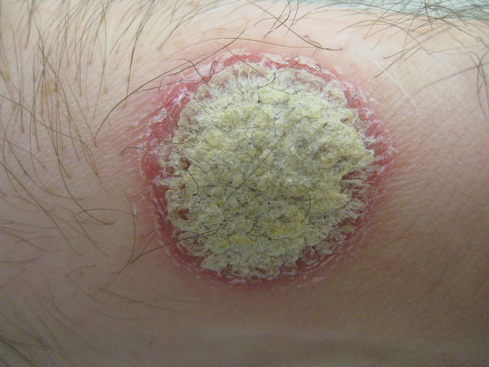
Psoriatic plaque, showing a silvery center surrounded by a reddened border. Source: Wikimedia. By James Heilman, MD – Own work, CC BY-SA 3.0

Considered to be the trigger for psoriais, the immune messenger molecule Interleukin-12 (IL-12) has now been shown to actually cause the skin disease but in fact protects against it. This finding also explains why common psoriasis drugs that block the messenger show insufficient treatment efficacy.

Psoriasis is a chronic inflammatory autoimmune disease that manifests as red, scaly skin patches. No causal treatment for the disease exists, but the symptoms can be significantly alleviated with modern therapies. The development of the skin disease arises from complex changes immune cell networks and the messengers they use for communication. Clinical trials showed that newly developed drugs that blocked only IL-23 are more effective than previous treatments targeting both IL-23 and IL-12 in psoriasis patients, but why this was so was not known. Now, researchers at the University of Zurich (UZH) have uncovered the underlying molecular mechanisms.

From human and mouse studies, they found that various cell types in the skin are also equipped with receptors for IL-12. Not only the T cells of the immune system, but also keratinocytes, horn-forming skin cells that build up the epidermis, can thus recognise the messenger. In fact, the recognition of interleukin-12 by these skin cells was responsible for the protective effect of the messenger, as the researchers found out. “Interleukin-12 is essential for the normal, physiological function of keratinocytes. For example, it prevents the increased cell division observed in psoriasis,” explained group leader Sarah Mundt from the Institute of Experimental Immunology at UZH.

“These results surprised us, because so far drugs for the treatment of psoriasis also aim at blocking interleukin-12,” said immunology professor Burkhard Becher.

“Our findings indicate that blocking IL-12 is not advisable, and such drugs should therefore no longer be used to treat psoriasis patients,” advised first author  Pascale Zwicky, PhD student. Accordingly, psoriasis drugs should only block the messenger substance IL-23, but no longer IL-23 and -12 together.

The UZH researchers’ findings could be important for the treatment of other diseases. “The combined blocking of IL-23 and -12 is also used in the treatment of chronic inflammatory bowel diseases and psoriatic arthritis,” said Prof Becher. “In these diseases, the role of IL-12 has not yet been sufficiently studied. But here, too, a protective role of the messenger substance is possible.”

Source: University of Zurich

A New, Lasting Diabetes Treatment

Source: National Cancer Institute on Unsplash

Israeli researchers have come up with a novel approach to the treatment of type 2 diabetes, using an autograft of muscle cells engineered to take in sugar at increased rates.

The disease’s long-term complications include heart disease, strokes, retinal damage leading to blindness, kidney failure, and poor blood flow in the limbs that may result in amputations. Currently a combination of lifestyle changes, medication, and insulin injections are used to treat it, however it is still associated with a 10-year reduction in life expectancy.

Professor Shulamit Levenberg led the study alomg with PhD student Rita Beckerman from the Stem Cell and Tissue Engineering Laboratory in the Technion’s Faculty of Biomedical Engineering. An autograft of muscle cells engineered to take in sugar at increased rates were tested in mice, which displayed normal blood sugar levels for months after a single procedure. The study findings were published in Science Advances.

Muscle cells are among the main targets of insulin, and they are supposed to absorb sugar from the blood. In their study, Prof. Levenberg’s group isolated muscle cells from mice and engineered these cells to present more insulin-activated sugar transporters (GLUT4). These cells were then grown to form an engineered muscle tissue, and finally put back into diabetic mice. The engineered cells not only proceeded to absorb sugar correctly, improving blood sugar levels, but also induced improved absorption in the mice’s other muscle cells through intercellular signalling. After this one treatment, the mice remained cured of diabetes for four months – the entire observation period. Their blood sugar levels remained lower, and they had reduced levels of fatty liver normally seen in type 2 diabetes.

Prof. Levenberg explained how the process worked. “By taking cells from the patient and treating them, we eliminate the risk of rejection.” These cells can easily integrate back into being part of the body and respond to the body’s signaling activity.

An effective treatment, especially as a once-off, could significantly improve both quality of life and life expectancy of those who have diabetes. The same method could also be used to treat various enzyme deficiency disorders.

Source: Technion Israeli Institute of Technology

Up to Five Times Higher Costs for Those with Rare Diseases

Credit: Thirdman on Pexels

By studying medical and insurance records indicates health care costs for people with a rare disease, researchers have found that these have been underestimated and are three to five times greater than the costs for people without a rare disease.

The findings, appearing in the Orphanet Journal of Rare Diseases, provides new evidence of the impact rare diseases could have on public health, suggesting that medical costs for individuals with rare diseases are on par with those for cancer and heart failure.  

“There needs to be greater public awareness of the large and growing medical footprint of rare diseases in society,” said senior author Anne Pariser, MD, director of the NCATS Office of Rare Diseases Research. “Only about 10% of rare diseases have an FDA-approved therapy for their treatment. The findings underscore an urgent need for more research, and earlier and more accurate diagnoses of and interventions for these disorders.”

Most of the 7000 to 10 000 known rare diseases disproportionately affect children, adolescents and young adults. Individually, most rare diseases might affect only a few hundred to a few thousand people around the world. Rare diseases however are collectively common, affecting an estimated 4% of the world’s population. Many of these diseases have a genetic cause, are serious or life-threatening and are hard to diagnose and treat.

The pilot study used International Classification of Diseases (ICD) codes, which designate a disease diagnosis and other methods, to determine those individuals with rare diseases and their direct medical costs for 14 rare diseases in four health care systems compared to non-rare disease patients of a similar age.

The 14 rare diseases represented a diverse set of disorders differing in prevalence, organ systems affected, age of onset, clinical course, and availability of an approved treatment or specific ICD code. These rare diseases include sickle cell disease, muscular dystrophy and eosinophilic esophagitis.

The analysis showed wide variations of rare diseases prevalence in the different health care systems, possibly due in part to geographic differences, as well as the use of public versus private insurance, which may include different patient group representation. Some genetic diseases can also have a higher prevalence in certain regions, due to demographic make-up.

With the Eversana health care system database, the cost per patient per year (PPPY) for those with a rare disease, ranged from $8 812 to $140 044 for rare diseases patients compared to $5862 for those without a rare disease. The NCATS data indicated PPPY costs ranging from $4859 to $18 994 for rare diseases patients versus $2211 for those without a rare disease.

Using patient medical records, the researchers also traced the diagnostic journeys of four people with a rare disease, including two individuals who had a form of Batten disease, an inherited neurological disorder, and two others with cystic fibrosis. These journeys provided detailed descriptions of direct medical costs, such as for hospitalisations and procedures for these diseases, and provided insights into patient clinical management before and after disease diagnosis.

Analysis of medical records also revealed that rare diseases patients often shared commanilities in symptoms (eg, seizures, infections, and developmental delay) and characteristics, which could aid in earlier diagnosis and treatmen. As many receive a rare disease diagnosis at a young age and because most rare diseases are serious conditions, rare disease patients are likely to require hospital time and incur greater medical expenses over a lifetime.

Such commonalities among rare disease patients could point to the potential use of machine learning techniques on health care system databases to improve diagnoses, said study co-author Joni L. Rutter, PhD, NCATS Acting Director.

The researchers would also like to determine whether these methodologies could be scaled to thousands of other known rare diseases.

“Ultimately, to improve the lives of people with rare diseases,” said Dr Rutter, “we need to find innovative ways, including new technologies, to help shorten the lengthy diagnostic odysseys so many patients and families experience and make more treatments available faster.”

Source: National Center for Advancing Translational Sciences

A New Clue to Disarming C. Difficile’s Toxic Weaponry

C difficile. Source: CDC

Therapeutic interventions for Clostridioides difficile infection (CDI) could make use of a glucosyltransferase domain (GTD) as an ideal molecular target, potentially yielding new, effective treatments for this deadly disease.

The study, published in Science Advancesprovided new insights into TcdB, the toxic molecular weaponry of C. difficile and its hypervirulent strains, creating an opportunity to disarm it.

CDI is the leading cause of antibiotic-associated diarrhoea and gastroenteritis-associated deaths worldwide, accounting for 500 000 cases and 29 000 deaths in the US every year and is classified by the Centers for Disease Control and Prevention as one of the top health threats. The emergence and spread of hypervirulent C. difficile strains is of global concern, resembling as it does the occurrence of new virus variants in current COVID pandemic. TcdB is one of two homologous C. difficile exotoxins, and TcdB alone is capable of causing the full spectrum of CDI diseases.

“We focused on the structure and function of TcdB’s crucial GTD, which is the toxin’s ‘warhead.’ The GTD is delivered by the toxin inside the host cells and causes most of the cytosolic damage to patients,” said corresponding author Rongsheng Jin, PhD, professor in the Department of Physiology & Biophysics at the UCI School of Medicine. “We discovered molecular mechanisms by which the GTD specifically recognises and blocks the physiological functions of the human GTPases Rho and R-Ras enzyme families that are crucial signaling molecules.”

The team also showed that the classic form of TcdB and the hypervirulent TcdB recognise their human targets in different ways, leading to distinct structural changes to the host cells caused by bacterial invasion.

“Once the GTD of TcdB is inside the cells, it is shielded by our cells and becomes inaccessible to passive immunotherapy. But our studies suggest that small molecule inhibitors could be developed to disarm the GTD, which will directly eliminate the root cause of disease symptoms and cellular damage,” Prof Jin explained. “This new strategy can potentially be integrated with and complement other CDI treatment regiments.”

Source: UCI School of Medicine

Gut Inflammation and Mental Health Link

Image by Falkurian Design on Unsplash

Using mouse models, scientists have discovered a link between gut inflammation and mental comorbidities. In response to gut inflammation like that caused by inflammatory bowel disease (IBD), they observed that the vascular barrier in the brain choroid plexus closes, clamping down access to the brain. The findings were published in Science.

Though this gut-brain vascular axis deregulation is likely a protective mechanism for the brain against inflammation, the findings suggest it may also result in the various cognitive and psychiatric symptoms that are occasionally associated with IBD.

Usually associated with intestinal inflammation, IBD can also cause a wide variety of symptoms in other organs. There is a robust link between anxiety and IBD; up to 40% of patients with IBD also present with psychiatric symptoms such as anxiety or depression. While the gut-brain axis is thought to be involved in driving these symptoms, no other related mechanisms are currently known.

Using a mouse model of intestinal inflammation, Sara Carloni and colleagues identified a potential pathogenic link between IBD and its associated mental comorbidities. According to the findings, the gut vascular barrier becomes more permeable due to the inflammatory process, which allows inflammation to spread beyond the intestines.

In response to this spread, the vascular barrier in the choroid plexus of the brain shuts down, which helps to protect the brain from inflammation. However, in doing so, the process also potentially impairs communications between organs and may hinder brain function.

In a mouse model of genetically driven closure of choroid plexus endothelial cells, Carloni and colleagues observed a deficit in short-term memory and anxiety-like behaviour. Thus, the mental deficits observed alongside IBD may result from deregulation of the gut-brain vascular axis, the authors said. This finding could be used for the development of therapeutic targets in treating some behavioural disorders.

Source: News-Medical.Net

Sleep is Also Important in Avoiding Overweight in Babies

Photo by Danijel Durkovic on Unsplash

While the importance has long been known, little research has examined the necessity of sufficient sleep during the first months of life. New research suggests that newborns who sleep longer and wake up less throughout the night are less likely to be overweight in infancy. Their results are published in Sleep.

“While an association between insufficient sleep and weight gain is well-established in adults and older children, this link has not been previously recognized in infants,” said study co-author Susan Redline, MD, MPH, senior physician in the Division of Sleep and Circadian Disorders at the Brigham. “In this study, we found that not only shorter nighttime sleep, but more sleep awakenings, were associated with a higher likelihood of infants becoming overweight in the first six months of life.”

To conduct this research, Dr Redline and colleagues observed 298 newborns born at Massachusetts General Hospital between 2016 and 2018. The infants’ sleep patterns were monitored using ankle actigraphy watches – devices that measure patterns of activity and rest over multiple days. Researchers obtained three nights’ worth of data at the one- and six-month marks while parents kept sleep diaries, recording their children’s sleep and wake episodes.

Scientists measured infant height and weight and determined their body mass index, classifying infants as overweight if they fell into or above the 95th percentile on the World Health Organization’s growth charts.

Notably, researchers found that just one extra hour of sleep was associated with a 26% decrease in overweight risk. Additionally, infants that woke up less during the night faced a lower risk of excess weight gain. Scientists speculate that having more sleep promotes routine feeding practices and self-regulation, factors that mitigate overeating.

Investigators noted an underrepresentation of African-American individuals and lower-income families in their dataset. Additionally, confounding variables, such as breastfeeding duration, could have impacted infant growth. In the future, the researchers aim to extend this study to evaluate how sleep patterns impact growth within the first two years of life and identify key factors that mediate the correlation between sleep and weight gain. They also aim to evaluate interventions for promoting healthy sleep habits.

“This study underscores the importance of healthy sleep at all ages,” said Dr Redline. “Parents should consult their pediatricians on the best practices to promote healthy sleep, like keeping consistent sleep schedules, providing a dark and quiet space for sleeping, and avoiding having bottles in bed.”

Source: EurekAlert!