Month: June 2021

Artificial Sweeteners Can Turn Gut Bacteria Bad

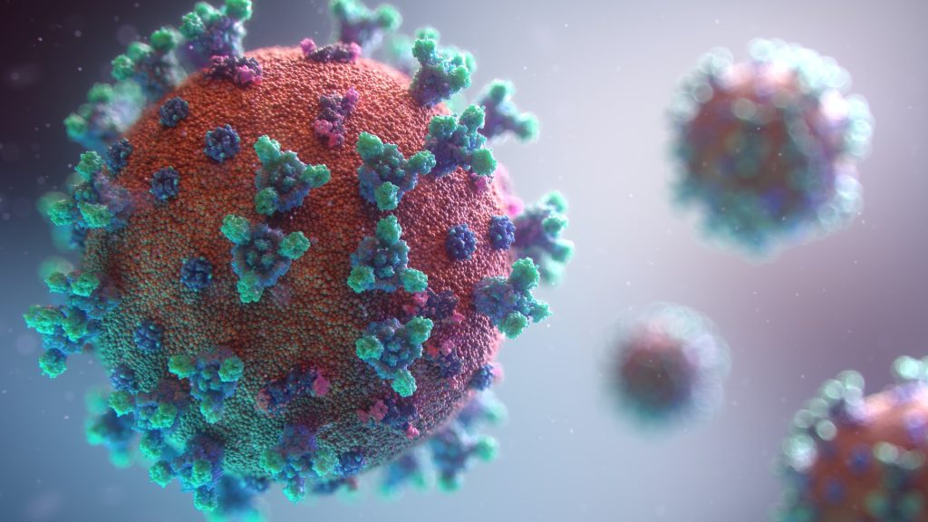
Source: Breakingpic on Pexels

Scientists have found that common artificial sweeteners can turn previously healthy gut bacteria pathogenic, invading the gut wall and potentially leading to serious health issues.

This study is the first to show the pathogenic effects of some of the most widely used artificial sweeteners (saccharin, sucralose, and aspartame) on two types of gut bacteria, Escherichia coli and Enterococcus faecalisE. faecalis is capable of crossing the intestinal wall to enter the bloodstream and congregate in the lymph nodes, liver, and spleen, causing a number of infections including septicaemia. To top it off, this commensal bacteria has emerged as a multi-drug resistant pathogen.

Previous studies have shown that artificial sweeteners can affect the composition of gut bacteria, but this new molecular research, led by academics from Anglia Ruskin University (ARU), has shown that sweeteners can also induce pathogenic features in certain bacteria. It found that these pathogenic bacteria can latch onto, invade and kill epithelial Caco-2 cells lining the intestinal wall.

This new study discovered that at a concentration equivalent to two cans of diet soft drink, all three artificial sweeteners significantly increased the adhesion of both E. coli and E. faecalis to intestinal Caco-2 cells, and differentially increased biofilm formation. Bacteria growing in biofilms are less sensitive to antimicrobial resistance treatment and are more likely to secrete toxins and express disease-causing virulence factors.

Additionally, all three sweeteners caused the pathogenic gut bacteria to invade Caco-2 cells found in the wall of the intestine, save for saccharin, which had no significant effect on E. coli invasion.

Senior author Dr Havovi Chichger, Senior Lecturer in Biomedical Science at ARU, said: “There is a lot of concern about the consumption of artificial sweeteners, with some studies showing that sweeteners can affect the layer of bacteria which support the gut, known as the gut microbiota.

“Our study is the first to show that some of the sweeteners most commonly found in food and drink—saccharin, sucralose and aspartame—can make normal and ‘healthy’ gut bacteria become pathogenic. These pathogenic changes include greater formation of biofilms and increased adhesion and invasion of bacteria into human gut cells.

“These changes could lead to our own gut bacteria invading and causing damage to our intestine, which can be linked to infection, sepsis and multiple-organ failure.

“We know that overconsumption of sugar is a major factor in the development of conditions such as obesity and diabetes. Therefore, it is important that we increase our knowledge of sweeteners versus sugars in the diet to better understand the impact on our health.”
Source: EurekAlert!

Journal reference: Shil, A & Chichger, H (2021) Artificial Sweeteners Negatively Regulate Pathogenic Characteristics of Two Model Gut Bacteria, E. coli and E. faecalis. International Journal of Molecular Sciences. doi.org/10.3390/ijms22105228.

The Origin Mystery of SARS-CoV-2 Deepens

SARS-CoV-2 viruses emerging from a human cell. Credit: NIAID

Australian researchers studying SARS-CoV-2 have discovered that the virus is most ideally adapted to infect human cells — instead of bat or pangolin cells, prompting renewed questions about its origin.

The scientists, from Flinders University and La Trobe University, described how they used high-performance computer modelling of SARS-CoV-2’s structure at the beginning of the pandemic to predict its ability to infect humans and a range of 12 domestic and exotic animals.

They were hoping to identify an intermediate animal vector that may have played a role in transmitting a bat virus to humans, and to understand any risk posed by the susceptibilities of pets and livestock.

Using genomic data from 12 animal species, the researchers painstakingly built computer models of the key ACE2 protein receptors for each species. These models were then used to calculate how strongly the SARS-CoV-2 spike protein bound to each species’ ACE2 receptor.

Surprisingly, the results showed that SARS-CoV-2 bound to ACE2 on human cells more tightly than any of the tested animal species, including bats and pangolins. If one of the animal species tested was the origin, it would normally be expected to show the highest binding to the virus.

“Humans showed the strongest spike binding, consistent with the high susceptibility to the virus, but very surprising if an animal was the initial source of the infection in humans,” said Professor David Winkler at La Trobe University.

The findings, originally released on the ArXiv preprint server, have now been peer reviewed and published in Scientific Reports.

“The computer modelling found the virus’s ability to bind to the bat ACE2 protein was poor relative to its ability to bind human cells. This argues against the virus being transmitted directly from bats to humans. Hence, if the virus has a natural source, it could only have come to humans via an intermediary species which has yet to be found,” says Flinders affiliated Professor Nikolai Petrovsky.

The team’s computer modelling also showed fairly strong binding of SARS-CoV-2 to ACE2 from pangolins, which are occasionally used as food or in traditional medicines. Professor Winkler noted that pangolins displayed the highest spike binding energy of all the animals in the study – significantly higher than bats, monkeys and snakes.

“While it was incorrectly suggested early in the pandemic by some scientists that they had found SARS-CoV-2 in pangolins, this was due to a misunderstanding and this claim was rapidly retracted as the pangolin coronavirus they described had less than 90% genetic similarity to SARS-CoV-2 and hence could not be its ancestor,” Prof Petrovsky said.

Similarity in spike proteins

As shown in this and other studies, the specific part of the pangolin coronavirus spike protein that binds to ACE2 was almost identical to its SARS-CoV-2 counterpart.

“This sharing of the almost identical spike protein almost certainly explains why SARS-CoV-2 binds so well to pangolin ACE2. Pangolin and SARS-CoV-2 spike proteins may have evolved similarities through a process of convergent evolution, genetic recombination between viruses, or through genetic engineering, with no current way to distinguish between these possibilities,” Prof Petrovsky said.

“Overall, putting aside the intriguing pangolin ACE2 results, our study showed that the COVID-19 virus was very well adapted to infect humans.”

“We also deduced that some domesticated animals like cats, dogs and cows are likely to be susceptible to SARS-CoV-2 infection too,” Prof Winkler added.

The question of how the virus came to infect humans currently has two main explanations. The virus may have jumped to humans from bats through an intermediary animal which remains to be identified. The other explanation making headlines in the media is an accidental release from a virology lab, where it perhaps was created in ‘gain of function‘ tests, which are carried out around the world to better understand pathogens. A number of organisations and governments, including the World Health Organization and the United States have urged further investigation to find out which of these is correct — though a definitive answer may take years.
How and where the SARS-CoV-2 virus adapted to become such an effective human pathogen remains a mystery, the researchers concluded, adding that finding the origins of the disease will help efforts to protect humanity against future coronavirus pandemics.

Source: EurekAlert!

Journal information: Sakshi Piplani et al, In silico comparison of SARS-CoV-2 spike protein-ACE2 binding affinities across species and implications for virus origin, Scientific Reports (2021). DOI: 10.1038/s41598-021-92388-5

New Machine Learning Tools Could Save Teeth

Photo by Kevin Bation on Unsplash

Machine learning tools could help identify those at greatest risk for tooth loss and refer them for further dental assessment for early interventions to avert or delay the conditions.

The study by researchers at the Harvard School of Dental Medicine compared five different algorithms using various combinations of variables to screen for risk. The results showed those that factored medical characteristics and socioeconomic variables, including race, education, arthritis, and diabetes, outperformed algorithms that relied on dental clinical indicators alone.

“Our analysis showed that while all machine-learning models can be useful predictors of risk, those that incorporate socioeconomic variables can be especially powerful screening tools to identify those at heightened risk for tooth loss,” said study lead investigator Hawazin Elani, assistant professor of oral health policy and epidemiology at HSDM.

The approach could be used to screen people globally and in a variety of health care settings even by non-dental professionals, she added. This approach could be applied around the world, even allowing non-dental professionals to screen patients.

Tooth loss can affect quality of life, well-being, nutrition, and social interactions. It is also associated with dementia. If the earliest signs of dental disease are identified, then the process can be delayed or averted with prompt treatment. However, many people with dental disease may not see a dentist until the process is too far gone. This is where screening tools could help identify those at highest risk and refer them for further assessment, the team said.

For the study, the researchers used data on nearly 12 000 adults from the National Health and Nutrition Examination Survey to design and test five machine-learning algorithms and assess their predictions for both complete and incremental tooth loss among adults based on socioeconomic, health, and medical characteristics.

A key point is that algorithms were designed to assess risk without a dental exam, though anyone at risk for tooth loss would still need one. The study’s findings point to the importance of socioeconomic factors.

“Our findings suggest that the machine-learning algorithm models incorporating socioeconomic characteristics were better at predicting tooth loss than those relying on routine clinical dental indicators alone,” Elani said. “This work highlights the importance of social determinants of health. Knowing the patient’s education level, employment status, and income is just as relevant for predicting tooth loss as assessing their clinical dental status.”

Low socioeconomic status populations have long been known to have greater rates of tooth loss, likely due to lack of regular access to dental care, among other reasons, the team said.

“As oral health professionals, we know how critical early identification and prompt care are in preventing tooth loss, and these new findings point to an important new tool in achieving that,” said Jane Barrow, associate dean for global and community health and executive director of the Initiative to Integrate Oral Health and Medicine at HSDM. “Dr. Elani and her research team shed new light on how we can most effectively target our prevention efforts and improve quality of life for our patients.”

Source: Harvard Medical School

Journal information: Hawazin W. Elani et al, Predictors of tooth loss: A machine learning approach, PLOS ONE (2021). DOI: 10.1371/journal.pone.0252873

Double Threat of Flu and S. Pneumoniae Unravelled

Streptococcus pneumoniae bacteria. Image by CDC on Unsplash

Researchers have found a further reason for why flu and Streptococcus pneumonia are such a deadly combination, by a surface protein causing it to stick to dead or dying lung cells. The finding by University of Alabama at Birmingham (UAB) follows thirty years after the discovery of the surface protein, called pneumococcal surface protein A, or PspA.

This new mechanism had been overlooked because it facilitates bacterial adherence only to dead or dying lung epithelial cells, not to living cells. Previously, researchers typically used healthy lung cell monolayers to search for bacterial adhesins that aid infection. In flu, the virus killing off lung cells was found to set the stage for S. pneumonia attachment to the airway, thereby worsening disease and pneumonia.

Study leaders Carlos Orihuela, PhD, and David Briles, PhD, professor at UAB, said their findings provide further explanation for how an infection by influenza A flu virus — followed by S. pneumoniae superinfection — causes severe pneumonia and a high death rate. Understanding of this mechanism could also lead to improvements for disease treatment and vaccination.

A historical example of the deadly synergy of flu infection followed by S. pneumoniae superinfection is found in banked lung samples from the 1918 Spanish influenza pandemic that killed 40 million to 50 million people — the vast majority of these samples showed co-infection or secondary infection with S. pneumonia.

The UAB research on PspA began with puzzling results from experimental lung infections of mice with influenza A, followed by either wild-type S. pneumonia that has the intact PspA gene, or a mutant S. pneumoniae that lacks PspA. Lung homogenates from mice infected with the wild-type had much higher numbers of S. pneumonia bacteria than lungs infected with the mutant. However, when researchers washed the interiors of the lungs and collected that bronchoalveolar lavage fluid, they counted similar numbers of the wild-type S. pneumonia and the mutant.

“This unexpected result was interpreted to mean that wild-type S. pneumoniae were more resistant to dislodgement than S. pneumonia with a pspA gene deletion, and it served as rationale for further experimentation,” Dr Orihuela said.

From this, the researchers were then able to show that PspA functions as an adhesin to dying host cells, as well as its previously established virulence mechanisms. The researchers also detailed the molecular mechanism of this bacterial adherence.

Both influenza A infection and release of the S. pneumoniae toxin pneumolysin cause death of lung epithelial cells. As they are dying, cells’ phosphatidylserine residues wind up on the outer cell membrane, where they bind the host enzyme glyceraldehyde-3-phosphate dehydrogenase, or GAPDH. In turn, the S. pneumoniae PspA on the bacteria surface binds to the GAPDH. PspA-GAPDH-mediated binding to lung cells increased S. pneumoniae localisation in the lower airway, and this was enhanced by pneumolysin exposure or co-infection with influenza A virus.

One of the fragments of protein responsible for the binding was introduced into the lungs of influenza-infected mice and reduced the disease severity of S. pneumoniae superinfection, presumably through binding competition.

“Our findings support the targeting of regions of PspA for therapeutic and vaccine development against influenza A/Streptococcus pneumoniae superinfections,” Dr Orihuela said. “Importantly, and despite more than 30 years since its discovery, PspA was not previously shown to function as an adhesin. Thus, our finding of PspA’s role in adherence substantially advances our knowledge on the interactions of S. pneumoniae with its host.”

Source: University of Alabama at Birmingham

Journal information: Sang-Sang Park et al, Streptococcus pneumoniae binds to host GAPDH on dying lung epithelial cells worsening secondary infection following influenza, Cell Reports (2021). DOI: 10.1016/j.celrep.2021.109267

Encouraging News for Migraine Sufferers

Photo by Usman Yousaf on Unsplash

According to a new study from the University of Toronto, 63% of Canadians who suffer from migraine headaches are able to flourish, despite the painful condition.

“This research provides a very hopeful message for individuals struggling with migraines, their families and health professionals. The findings of our study have contributed to a major paradigm shift for me. There are important lessons to be learned from those who are flourishing,” said lead author Esme Fuller-Thomson, Professor at both the Factor-Inwentash Faculty of Social Work and the Department of Family & Community Medicine, University of Toronto.

Over the past decade, Prof Fuller-Thomson has published on negative mental health outcomes associated with migraines, including suicide attempts, anxiety disorders and depression.

Migraine is a complex,   genetically influenced disorder characterised by episodes of moderate-to-severe headache, usually unilateral and associated with nausea and increased sensitivity to light and sound. A migraine headache is the seventh most disabling disorder in the world. However, there is little research investigating factors associated with mental health and well-being among sufferers.

The University of Toronto study investigated optimal mental health in a nationally representative sample of 2186 Canadian adults diagnosed with migraines. The data were drawn from Statistics Canada’s Canadian Community Health Survey-Mental Health. To be defined in excellent mental health, respondents had to achieve three things: 1) almost daily happiness or life satisfaction in the past month, 2) high levels of social and psychological well-being in the past month, and 3) freedom from generalised anxiety disorder and depressive disorders, suicidal thoughts and substance dependence for at least the preceding full year.

“We were so encouraged to learn that more than three in every five migraineurs were in excellent mental health and had very high levels of well-being,” said Prof Fuller-Thomson.

Those experiencing migraines who had at least one person they could confide in were four times more likely to be in excellent mental health than those without. Additionally, those using religious or spiritual beliefs to cope with everyday difficulties had 86% higher odds of excellent mental health than those who did not use spiritual coping. Poor physical health, functional limitations, and a history of depression were also found to inhibit excellent mental health among those with migraines.

“Health professionals who are treating individuals with migraines need to consider their patients’ physical health needs and possible social isolation in their treatment plans” said co-author Marta Sadkowski, a recent nursing graduate from the University of Toronto.

Source: EurekaAlert!

Journal reference: Fuller-Thomson, E & Sadkowski, M., (2021) Flourishing Despite Migraines: A Nationally Representative Portrait of Resilience and Mental Health among Canadians. Annals of Headache Medicine. doi.org/10.30756/ahmj.2021.05.02.

Intense Training Results in Temporary Mitochondrial Impairment

Photo by Quino Al on Unsplash

Elite athletes have temporary mitochondrial impairment following intense workouts, according to new research, which suggests they may need to be mindful about overtraining. 

Mitochondria are organelles that are responsible for most of the useful energy derived from the breakdown of carbohydrates and fatty acids, which is converted to ATP by the process of oxidative phosphorylation. Mitochondrial capacity is a term used to describe the body’s ability to generate energy, and is one factor associated with increased athletic performance during endurance exercise. Previous research found that untrained recreational athletes had a decrease in mitochondrial capacity after sprinting exercises.

In this study, the researchers worked with a small group of male elite athletes, many of whom were national title holders or had international recognition for their performance in cycling and triathlon. The athletes participated in a four-week training programme in their primary sport, which consisted of two to four days of low-to-moderate–intensity endurance workouts, followed by three days of more intense training. These intense workouts included high-intensity interval training in the morning, followed by a seven-hour break and then a moderate-intensity cycling session in the afternoon. Each volunteer did between 12 and 20 hours of activity per week. The athletes, though used to heavy training, were not accustomed to this specific workout schedule.

The researchers were surprised to observe that the highly trained participants’ mitochondrial capacity was impaired after the month-long training period. “We thought that elite athletes should be more resistant against [these] kind of alterations,” said Filip Larsen, PhD, of the Swedish School of Sport and Health Sciences and corresponding author of the study.

However, elite athletes may be able to prevent temporary mitochondrial impairment by listening to their bodies, the researchers suggested. By paying attention to changes such as “mood disturbances, reductions in maximal heart rate [during exercise] and muscles that feel heavy and unresponsive” top athletes may be able to pull back and avoid overtraining situations that could contribute to reduced mitochondrial content and function, Larsen explained. “Exercise is good for you, but too much unaccustomed training might have mitochondrial consequences.”

The study also found that reduced mitochondrial capacity did not affect exercise performance, suggesting that oxygen delivery from the heart to the muscles plays a more important role than mitochondrial function in performance. Expression of three proteins with strong antioxidant properties were also found to be increased in the muscles after intense training.

Source: American Physiological Society

Journal information: Daniele A. Cardinale et al, Short term intensified training temporarily impairs mitochondrial respiratory capacity in elite endurance athletes, Journal of Applied Physiology (2021). DOI: 10.1152/japplphysiol.00829.2020

Positivity Rate at 25% as Lockdown Upgrades Expected

President Cyril Ramaphosa is expected to meet with the National Coronavirus Command Council (NCCC) to discuss the government’s response to the third COVID wave, which includes the possibility of new restrictions. 

Several bodies have strongly urged upgrading to a harder lockdown, including the South African Medical Association, the Gauteng Provincial Government, medical professionals, and now the Ministerial Advisory Committee on Covid-19.

Earlier this week Ramaphosa indicated that the government will have to increase its COVID containmant measure – especially in Gauteng province. He noted that the country’s first hard lockdown in March 2020, one of the strictest in the world, did help cut infection rates at the start of the pandemic.

South Africa recorded 17 493 new cases, a new daily high for the third wave, of which 10 806 were in Gauteng. Case positivity rate increased to 24.92%. A report released on Wednesday by the South African Medical Research Council showed that 1349 excess deaths in Gauteng for the week ending 13 June, of which 431 were due to COVID/

Warnings and failure to act

In an interview with The Money Show with Bruce Whitfield this Monday, Netcare CEO Richard Friedland had warned that the numbers of Covid-19 patients “are overwhelming facilities at the moment”.

Since Wednesday last week, Gauteng’s hospitals had been battling with a “mass casualty situation” , not unlike the aftermath of a train accident, or the collapse of a sports stadium, with “injuries on a massive scale”. But, with COVID, he said, the crisis is not over in a couple of hours, but remains ongoing.

With no evidence of a peak in case numbers, Friedland said that, “I’m afraid that these numbers are demonstrating that [without] a Level 5 lockdown in Gauteng, we may not see the end of this surge for some time.”

Professor Koleka Mlisana, co-chairperson of the Ministerial Advisory Committee on Covid-19, says that tighter restrictions are likely needed to help curb infections.

Prof Mlisana said that the other major crisis is making sure that there are sufficient hospital beds in Gauteng. This includes additional facilities, staffing members and beds to ensure the system is not overwhelmed, she said.

Prof Mlisana said that this was down to a lack of preparation by the government, despite warnings from the advisory committees. 

Source: BusinessTech

WHO Urges Equitable Travel Requirements

Photo by Tim Gouw on Unsplash

The WHO has urged that as air travel is restored, vaccinations should not be a prerequisite for travellers, potentially locking out those in poorer regions, especially Africa.

In a virtual press briefing on Thursday, Dr Matshidiso Moeti, World Health Organization Regional Director for Africa said that the WHO believes that schemes to remove quarantine and entry restrictions for travellers that have been vaccinated, are discriminatory and could deepen already existing inequalities even further.

Meanwhile, she warned that Africa’s third wave, already underway in 12 countries, with cases rising in another 14, threatens to be the worst yet with 5.3 million cases across the continent. It is projected that in three weeks the third wave will surpass the previous wave’s peak.

Public fatigue and new variants are driving this surge across Africa, with Delta the variant  detected in 14 countries. She stated that Africa can “blunt this third wave” but “the window of opportunity is closing”.

The WHO aims to strengthen variant surveillance in Africa by reinforcing the regional laboratory hub have a 8 to 10 fold increase in next 6 months for genome sequencing

Though vaccination rates remain low in Africa, there is nevertheless a great demand for vaccines, with 18 countries having used over 80% of the vaccines received through COVAX. Fortunately only mild side effects from the vaccines have been seen in African communities, she said.

Mr Kamil Alawadi, Regional Vice President for Africa and Middle East, International Air Transport Association (IATA) said that inconsistent requirements added additional complications in travel, increasing cost for the passenger and the airline. For travellers, PCR testing can range from $100 up to $400 for a single, one direction trip.

The key requirement for the recovery of the airline industry is the lifting of restrictions, said Alwadi, citing a survey that showed that 84% of passengers will not fly if there were quarantines in place. However, demand still existed for air travel, as evidenced by travel bookings spiking as soon as governments relaxed their border restrictions.

Alawadi said that the IATA agreed with the WHO that only lifting quarantine requirements for vaccine individuals was inequitable, and that “a robust and flexible testing system” was needed in place of quarantine, using systematic testing at the point of departure such as rapid antigen tests which are cheaper, faster and more accessible.

Graphic from Skyscanner.net showing countries with major travel restrictions from South Africa (red, 83 countries), moderate (orange, 29) and low restrictions (green, 42)

The situation was urgent for the African aviation industry as it had lost USD7.8 billion in 2020, with eight airlines filing for bankruptcy, he noted. This was against a background of USD430 billion global loss for the industry, though he noted that some countries are seeing a rebound to 2019 numbers for domestic travel. However, it is projected that losses will only stop by 2023 and return to profit by 2024.

The IATA has developed protocols in concert with the  International Civil Aviation Organization (ICAO) and WHO that will be non-discriminatory not require vaccinations, said Alwadi. However the aviation industry is sinking very rapidly without governmental support.

Cheaper Way of Monitoring Coronavirus Variants

Researchers at Karolinska Institutet in Sweden have developed a technology for cost-effective surveillance of the global spread of new SARS-CoV-2 variants.  This could help low- and middle-income countries monitor variants in their own borders.

From the beginning of the pandemic, thousands of viral genomes have been sequenced in order to reconstruct the evolution and global spread of the coronavirus. Dependent on these is the identification of particularly concerning variants.

To achieve global surveillance of the SARS-CoV-2 genome, the sequencing and analysis of numerous samples cost-effectively is key. Therefore, researchers in the Bienko-Crosetto laboratory at Karolinska Institutet and Science for Life Laboratory (SciLifeLab) in Sweden have developed a new method, COVseq, that can be used for surveillance of the viral genome on a massive scale at a low cost.

Multiplex PCR (polymerase chain reaction) is used to make more copies of the virus. The samples are then labeled and pooled together in the same sequencing library, using a previous method developed in their laboratory and now adapted for SARS-CoV-2 analysis.

“By performing reactions in very small volumes and pooling together hundreds of samples into the same sequencing library, we can sequence potentially thousands of viral genomes per week at a cost of less than 15 dollars per sample,” said co-first author Ning Zhang, postdoctoral researcher at the Department of Medical Biochemistry and Biophysics, Karolinska Institutet.

Comparative analyses of 29 SARS-CoV-2 positive samples revealed that COVseq could detect small changes in the genome as well as standard methods. Analysing 245 additional samples, they showed that COVseq could also detect emerging variants of concern well. COVseq’s key advantage over existing methods is cost-effectiveness.

“Our inexpensive method could immediately be used for SARS-CoV-2 genomic surveillance by public health agencies and could also be easily adapted to other RNA viruses, such as influenza and dengue viruses,” said last author Nicola Crosetto, senior researcher at the Department of Medical Biochemistry and Biophysics, Karolinska Institutet.

Source: Karolinska Institutet

Journal information: COVseq is a cost-effective workflow for mass-scale SARS-CoV-2 genomic surveillance. Nature Communications, 23 June 2021, DOI: 10.1038/s41467-021-24078-9

Inability to Dispose of Old Cells Leads to Macular Degeneration

Photo by v2osk on Unsplash

Researchers have discovered that failing to dispose of old photoreceptor cells leads to age-related macular degeneration.

The estimated number of people worldwide with age-related macular degeneration in 2020 was 196 million, increasing to 288 million in 2040. Though more than 50 genes are associated with the condition, the precise mechanism is unknown. Most people have a form of the condition, for which there are no known effective treatments.

In order to develop new therapies to treat the disease, University of Maryland School of Medicine (UMSOM) researchers are starting to understand what goes wrong in the disease. Using human and mice tissue, researchers showed that the process which removes the eye’s old, damaged light sensors is disrupted in macular degeneration.

Previously, the lead researcher had found out that many families with hearing disorders had genetic mutations in the gene for the CIB2 protein, and later work also showed that CIB2 was needed for vision in a large human family, as well as in zebrafish. Now, in this latest study, his team built on that previous work to dissect the intricate cell mechanisms behind retinal degeneration.

The team compared healthy mouse eyes to those from a mouse with the CIB2 protein genetically deleted. These CIB2 mutant mice were not disposing of their old light sensor proteins, called photoreceptors, like healthy mouse eyes did.  

“Photoreceptors continue growing in tiny columns in the eye, but over time, light damages the photoreceptors. To combat this, support cells in the eye slowly munch on the old, damaged photoreceptors, keeping the columns the correct length,” explained first author Saumil Sethna, PhD, at the University of Maryland School of Medicine. “If the photoreceptors are not removed, or if the process is backed up due to slow digestion by the support cells, like in the CIB2 mutant mice, the undigested material builds up over time, which may contribute to blindness.”

The researchers then identified several components in this photoreceptor recycling process, including a group of proteins collectively called mTORC1, which is involved in many human diseases, including cancer, obesity, and epilepsy.

Since mTORC1 (part of a family called mTOR) is a decision-maker for many cellular functions including cleaning up cellular debris, the researchers examined mTORC1’s activity in the CIB2 mutant mice and saw that it was overactive. mTORC1 was also found to be overactive in eye tissue of people with a form of age-related macular degeneration. The findings therefore indicate that drugs against mTORC1 may be effective treatments for the most common type of age-related macular degeneration, according to the researcher.
“Researchers have tested many small molecules directed at mTORC1 to treat various diseases, but the problem is that mTOR is needed for so many other cell functions that there are major side-effects when you tinker with it,” said senior author Zubair M Ahmed, PhD, Professor of Otorhinolaryngology-Head & Neck Surgery and Ophthalmology at the University of Maryland School of Medicine. “In our study, we found a backdoor way to regulate mTORC1, which may bypass many of the unpleasant side-effects that normally occur with suppressing mTORC1. We think we may be able to use our new knowledge of this mechanism to develop treatments for age-related macular degeneration and other diseases as well.”

Source: University of Maryland School of Medicine