Day: June 29, 2021

The Emerging Treatment-resistant Fungus Threat

Professor Rodney E. Rohde, a public health and clinical microbiology expert at Texas State University, warned in article for The Conversation of the growing threat of fungal resistance — a problem drawing much less attention than antibiotic resistance. 

 Athlete’s foot, thrush, ringworm and other ailments are caused by fungi, and some are serious risks to health and life. Among these is Candida auris, a pathogenic fungus. Fungi generally have not caused major disease, so there is a lack of funding in this area and there are limited antifungal agents that can treat C. auris.

Most fungal infections around the world are caused by the genus Candida, particularly the species called Candida albicans. But there are others, including Candida auris, which gets its name ‘auris’, Latin for ear, because it was first identified from an external ear canal discharge in 2009.

Candida normally lives on the skin and inside the body, such as in the mouth, throat, gut and vagina, without causing any problems. It exists as a yeast and is thought of as normal flora, harmless microbes. However when the body is immuno-compromised, these fungi become opportunistic pathogens, something happening around the world with multidrug-resistant C. auris.

The threat of Candida auris

C. auris infections, or fungaemia, have been reported in 30 or more countries. They are often found in the blood, urine, sputum, ear discharge, cerebrospinal fluid and soft tissue, and occur in people of all ages. According to the US Centers for Disease Control, the mortality rate in the US has been reported to be between 30% to 60% in many patients who had other serious illnesses. In a 2018 review of research on the global spread of the fungus, researchers estimated mortality rates of 30% to 70% in C. auris outbreaks among critically ill patients in intensive care.

Recent surgery, diabetes and broad-spectrum antibiotic and antifungal use are risk factors. Furthermore, immuno-compromised patients are at greater risk than those with healthy immune systems.

C. auris can be difficult to identify with conventional microbiological culture techniques, which leads to frequent mis-identification and under recognition. This yeast is also known for its tenacity to easily colonise the human body and environment — including medical devices. People in nursing homes and patients with catheters, on ventilation etc seem to be at highest risk.

The CDC has set C. auris infections at an “urgent” threat level because 90% are resistant to at least one antifungal, 30% to two antifungals, and there are some resistant to all three available classes of antifungals. This multidrug resistance has led to outbreaks in health care settings, especially hospitals and nursing homes, that are extremely difficult to control.

The double threat of COVID and C. auris

For hospitalised COVID patients, antimicrobial-resistant infections may be a particularly devastating risk. The mechanical ventilators often used to treat serious COVID are breeding grounds and highways for entry of environmental microbes like C. auris. Further, according to a September 2020 paper, hospitals in India treating COVID have detected C. auris on surfaces including “bed rails, IV poles, beds, air conditioner ducts, windows and hospital floors.” The researchers termed the fungus a “lurking scourge” amid the COVID pandemic. Termed ‘white fungus’, these fungal infections typically arise a week to 10 days after being in the ICU.

The same authors reported in a November 2020 CDC article that of 596 COVID-confirmed patients in a New Delhi ICU from April 2020 to July 2020, 420 patients required mechanical ventilation. Of these, 15 were infected with candidemia fungal disease and eight of those infected (53%) died. Ten of the 15 patients were infected with C. auris; six of them died (60%).

How to deal with this?

With fewer and fewer antifungal options,  CDC is recommending a focus on preventing C. auris infections. This involves better hand hygiene and improving infection prevention and control in medical care settings, judicious and thoughtful use of antimicrobial medications, and stronger regulation limiting the over-the-counter availability of antibiotics.

Source: The Conversation

Journal information: Anuradha Chowdhary et al, The lurking scourge of multidrug resistant Candida auris in times of COVID-19 pandemic, Journal of Global Antimicrobial Resistance (2020). DOI: 10.1016/j.jgar.2020.06.003

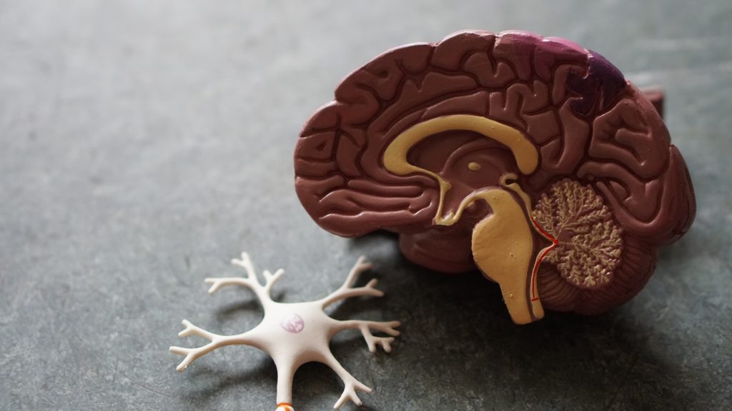
Are We Wrong About Amyloid Plaques in Alzheimer’s?

A recent study sheds new light on the disease and the highly debated aducanumab, a new drug recently approved by the FDA that treats the amyloid plaques.

Led by the University of Cincinnati and conducted in collaboration with the Karolinska Institute in Sweden, the study claims that the treatment of Alzheimer’s disease might lie in normalising levels of a brain protein called amyloid-beta peptide. This protein is needed in its original, soluble form to keep the brain healthy, but it sometimes hardens into ‘brain stones’ or clumps, called amyloid plaques.

“It’s not the plaques that are causing impaired cognition,” said senior author Alberto Espay, professor of neurology at UC. “Amyloid plaques are a consequence, not a cause,” of Alzheimer’s disease, stated Prof Espay, who is also a member of the UC Gardner Neuroscience Institute.

Since its discovery, scientists have focused on treatments to eliminate the plaques. But the UC team, he said, viewed it differently: Cognitive impairment could be due to a decline in soluble amyloid-beta peptide instead of the corresponding accumulation of amyloid plaques. 
To test their hypothesis, they analyzed the brain scans and spinal fluid from 600 individuals enrolled in the Alzheimer’s Disease Neuroimaging Initiative study, who all had amyloid plaques. From there, they compared the amount of plaques and levels of the peptide in the individuals with normal cognition to those with cognitive impairment. They found that individuals with high levels of the peptide were cognitively normal, despite the numbers of plaques in their brains.

They also found that higher levels of soluble amyloid-beta peptide were associated with a larger hippocampus, the area of the brain most important for memory.

According to the authors, as we age most people develop amyloid plaques, but few people develop dementia. In fact, by the age of 85, 60% of people will have these plaques, but only 10% develop dementia.

“The key discovery from our analysis is that Alzheimer’s disease symptoms seem dependent on the depletion of the normal protein, which is in a soluble state, instead of when it aggregates into plaques,” said co-author Kariem Ezzat from the Karolinska Institute.

The most relevant future therapeutic approach for the Alzheimer’s program would then be to restore these brain soluble proteins to their normal levels, said Prof Espay.

The research team is now working to test their findings in animal models. If successful, future treatments may be very different from those tried over the last two decades. Treatment, says Espay, may consist of increasing the soluble version of the protein in a manner that keeps the brain healthy while preventing the protein from hardening into plaques.  

Source: University of Cincinnati 

Journal information: Andrea Sturchio et al, High cerebrospinal amyloid-β 42 is associated with normal cognition in individuals with brain amyloidosis, EClinicalMedicine (2021). DOI: 10.1016/j.eclinm.2021.100988

Connective Tissue Protein Has Immune Role

Photo by National Cancer Institute on Unsplash

A new study finds that a connective tissue protein also encourages immune responses that fight bacterial infections, while restraining responses that can be deadly in sepsis.

The study focuses on the extracellular matrix (ECM) of connective tissues, once viewed merely as structural material. It is now increasingly recognised as a signaling partner with nearby cells in normal function, as well as being involved in disease. Fibroblasts are important players in the ECM; these cells make tough structural matrix proteins like collagen. The study was published online June 28 in the Proceedings of the National Academy of Sciences.

The new analysis found that lumican, a protein-sugar combination (proteoglycan) secreted by fibroblasts, and known to partner with collagen in connective tissues, also promotes immune system responses in immune cells called macrophages that fight bacterial infections. The study also found that lumican protects tissues by holding back a different type of immune response that reacts to DNA, whether from an invading virus, or released from cell death.

Such inflammatory responses are a transition into healing, but in sepsis they grow out of control, causing damage to the body’s own tissues. Sepsis affects 48.9 million people worldwide, the authors said, but the ECM’s role the condition is largely unknown.

“Lumican may have a dual protective role in ECM tissues, promoting defense against bacteria on the one hand, and on the other, limiting immune overreactions to DNA that cause self-attack, or autoimmunity,” said corresponding study author Shukti Chakravarti, PhD, professor in the Department of Ophthalmology and the Department of Pathology at NYU Langone Health.

The findings suggest that connective tissue, and extracellular matrix proteins like lumican, usually operate outside of cells, but as disease or damage break down ECM, get sucked into and regulate immune cells homing in on the damage.

Lumican  interacts with two proteins on surfaces of immune cells that control the activity of toll-like receptors, which recognise structural patterns common to molecules made by invading microbes, said the researchers. As they are less specific than other parts of the immune system, toll-like receptors can also cause attacks by immune cells on the body’s own tissues if over-activated.

In this study, the researchers found that lumican promotes the ability of toll-like receptor (TLR)-4 on the surfaces of immune cells to recognise bacterial cell-wall toxins called lipopolysaccharides (LPS). Lumican, by attaching to two proteins, CD14 and Caveolin1, probably using collagen-covered regions, stabilises their interactions with TLR4 to increase its ability to react to LPS. This results in production of the signalling protein TNF alpha, which amplifies immune responses.

Along with describing the effect of lumican on the surfaces of immune cells, the new study finds that lumican is taken up from outside cells into membrane-bound pouches, called endosomes, and pulled into cells. Such compartments deliver ingested bacteria to other endosomes that destroy them, heighten inflammation, or produce protective interferon responses. Once pulled inside, the researchers found, lumican bolstered TLR4 activity by slowing down its passage into lysosomes, pockets where such proteins are broken down and recycled.

However, while it encouraged TLR4 activity on cell surfaces, lumican, once inside immune cells, had the opposite effect on toll-like receptor 9 (TLR9), which reacts to DNA instead of bacterial LPS.

Mice with the lumican gene deleted had trouble both fighting off bacterial infections (less cytokine response, slower clearance, greater weight loss), and trouble restraining the immune overreaction to bacteria (sepsis). Elevated lumican levels were also found in human sepsis patients’ blood plasma, and that human immune cells (blood monocytes) treated with lumican had elevated TLR4 activity but suppressed TLR9 responses.

“As an influencer of both processes, lumican-based peptides could be used as a lever, to tweak inflammation related to TNF-alpha, or endosomal interferon responses, to better resolve inflammation and infections,” suggested George Maiti, PhD, a postdoctoral fellow in Dr Chakravarti’s lab.

“Our results argue for a new role for ECM proteins at sites of injury. Taken up by incoming immune cells it shapes immune responses beyond the cell surface by regulating the movement and interaction of endosomal receptors and signaling partners,” said Dr Chakravarti.

Source: NYU Langone Health

Journal information: George Maiti et al., “Matrix lumican endocytosed by immune cells controls receptor ligand trafficking to promote TLR4 and restrict TLR9 in sepsis,” PNAS (2021). www.pnas.org/cgi/doi/10.1073/pnas.2100999118

Trauma Patients with COVID at Great Risk

Photo by Nate Isaac on Unsplash

The COVID pandemic has placed a great strain on healthcare resources, with a number of indirect impacts ranging from increased incidence of heart attacks to decreased cancer screenings, but also increased the risk of complications and death among trauma patients with COVID. 

The study revealed that the risk of death for COVID-positive patients in trauma centres across the US state of Pennsylvania was six times higher than non-COVID-negative patients with similar injuries. Complication risk in COVID-positive patients was doubled for venous thromboembolism, renal failure, need for intubation, and unplanned ICU admission, and was five times greater for pulmonary complications. In patients over age 65, the risks were even higher. The findings were recently published in The Journal of Trauma and Acute Surgery.  

“COVID had the largest impact on patients whose injuries were relatively minor, and who we would have otherwise expected to do well,” said lead author Elinore Kaufman, MD, MSHP, an assistant professor in the Division of Trauma, Surgical Critical Care and Emergency Surgery at Penn Medicine. “Our findings underscore how important it is for hospitals to consistently test admitted patients, so that providers can be aware of this additional risk and treat patients with extra care and vigilance.”

Researchers conducted a retrospective study of 15 550 patients admitted to Pennsylvania trauma centers from March 21, 2020, (when non-essential businesses statewide were ordered close) to July 31, 2020. Of the 15 550 patients, 8170 were tested for the virus, and 219 tested positive. During this period, the researchers evaluated length of stay, complications, and overall outcomes for patients who tested positive for COVID, compared to patients who did not have the virus. They found that rates of testing increased over time, from 34% in April 2020 to 56% in July. Centres had a great variability in testing, a median of 56.2% of the time with a range of 0 to 96.4%.

“First, we need to investigate how to best care for these high-risk patients, and establish standard protocols to minimise risks,” said senior author Niels D Martin, MD, chief of Surgical Critical Care and an associate professor in the division of Trauma, Surgical Critical Care and Emergency Surgery. “Second, we need more data on the risks associated with patients who present symptoms of COVID, versus those who are asymptomatic, so we can administer proven treatments appropriately and increase the likelihood of survival with minimal complications.”

Source: University of Pennsylvania

‘Far Too Many Oocytes’ Being Extracted in IVF

Photo by MART PRODUCTION from Pexels

A study has suggested that IVF clinics in the UK may be retrieving “far too many oocytes” and that most of them “may never be used and are probably discarded”, a finding that may well represent global practice.

Studies indicate that the optimal and safe number of oocytes needed for achieving an ongoing pregnancy is between six and 15. However, the use of egg freezing (such as to preserve fertility to a later age, known as social egg freezing), frozen embryo replacement (FER) cycles and aggressive stimulation regimes has raised this number in order to boost success rates in older women and in poor responders who produce fewer eggs. What is not known is the impact of numbers of eggs retrieved and of over-stimulation practices on the health of patients, and on their emotional state and finances.

Details of the analysis were presented online at the virtual Annual Meeting of ESHRE by Dr Gulam Bahadur from North Middlesex University Hospital, London.

More than 1.625 million eggs in the UK were retrieved from 147,274 women between 2015 and 2018. Although an average of 11 eggs was collected per patient, 16% of cycles were associated with 16-49 oocytes retrieved (per cycle) and 58 women each had over 50 eggs collected in a single egg retrieval procedure.

“Our observations suggests that the high oocyte number per retrieval procedure needs re-evaluation,” said Dr Bahadur. “In particular, this needs to focus on the side effects, including ovarian hyperstimulation syndrome and procedure-related complications, and on the fate of unused frozen oocytes and the costs associated with freezing them.

“Patients should be advised that it’s better to collect fewer eggs leading to good quality embryos which may go to term and result in a healthy baby.”

This report is based on all UK IVF clinics and relates to non-donor fertility treatment carried out between 2015 and 2018 during which 172 341 fresh oocyte retrieval cycles took place. All outcomes and patterns remained uniform over the four years.

A substantial number (n = 10 148) of cycles did not yield any oocytes. Over half (53%) of all IVF cycles were in the desired egg yield range of 6-15. In addition, a quarter of cycles yielded 1-5 eggs; 14% produced 16-25; and a minority (2%) resulted in 26-49 oocytes. The authors point out that multiple birth rates increase significantly from 6-15 oocytes onwards, which increases the risk of birth complications and low birth weight.

A total of 931 265 embryos resulted from all eggs retrieved – a fertilisation rate of 57%. Of the embryos created, more than one in five (22% or 209,080) were transferred into the uterus, while a slightly higher proportion (24% or 219, 563) were frozen.

The fate of the unfertilised oocytes (43%) is unknown, though they are usually discarded. Most of the embryos not transferred (54%) will likely be discarded after patients have paid for several years of storage.

“This comes with a financial and emotional cost,” said Bahadur. “Patients build an attachment with this frozen material and there’s insufficient counselling to support them. They should be given more information about the implications of freezing eggs and embryos.”

Source: European Society of Human Reproduction and Embryology

Medical Device Warning for Certain Apple Devices

Apple has released a list of its products that it advises should be kept a “safe distance” away from sensitive medical devices such as pacemakers and implanted defibrillators. These products are iPhone 12 models, Apple Watch and MacBook Pro. 

A number of consumer-electronic devices contain components, such as magnets, which are known to interfere with medical devices. A number of other manufacturers, for example Samsung and Huawei, have issued similar guidance for some of their products.

Heart health is a promoted feature of some Apple products; certain Apple Watches can make electrocardiogram tests and display the results to the user, as well as recording the data for later medical examination. A number of studies have shown that Apple watches can detect cardiovascular problems such as atrial fibrillation with a fairly high degree of sensitivity. However, the current notice warns of the risks posed by components in some products.

“Under certain conditions, magnets and electromagnetic fields might interfere with medical devices,” Apple wrote, noting “implanted pacemakers and defibrillators might contain sensors that respond to magnets and radios when in close contact”.

Implanted defibrillators send electrical pulses to regulate abnormal heart rhythms. Apple said the listed products should be kept more than 15cm away from medical devices, double that if they are wirelessly charging.

The support page that listed the devices, had said earlier this month that iPhone 12 models were “not expected to pose a greater risk of magnetic interference to medical devices” than other iPhones.

However, the website MacRumours, which first noted the list, pointed to research suggesting that the iPhone 12 could interfere with implanted devices.

A study published June 2 in the Journal of the American Heart Association found that “Apple’s iPhone 12 Pro Max MagSafe technology can cause magnet interference”, and so had the potential “to inhibit life-saving therapy”.

The researchers acknowledged the study’s small scale as a limitation, though in a press release lead investigator Dr Michael Wu wrote that they were surprised by the strength of the magnets in the iPhone 12.

“In general, a magnet can change a pacemaker’s timing or deactivate a defibrillator’s life-saving functions, and this research indicates the urgency for everyone to be aware that electronic devices with magnets can interfere with cardiac implantable electronic devices.”

However Marie Moe, a computer security consultant for Mnemonic, told the BBC she was not worried.

“These Apple gadgets are generally not emitting large magnetic fields, unlike heavy machinery, big concert speakers or welding equipment that anyone with a pacemaker should be more concerned about getting in close proximity to,” she said. She is a pacemaker user herself and studies their use.

Ms Moe added that magnets as strong as those in the iPhone 12 could only put the pacemaker into “a kind of safety mode where the pacing is constant”, which would revert back once the device was removed.

Jo Whitmore, senior cardiac nurse at the British Heart Foundation, agreed that devices kept at a safe distance were not cause for concern. “It’s perfectly OK to use a smartphone when you have a pacemaker, and they’re designed to return to normal settings once the magnet is moved away,” she said.

She added that patients should check the device instructions or talk to the manufacturer if they are concerned, and they could also contact their doctor or pacing clinic.

Source: BBC News