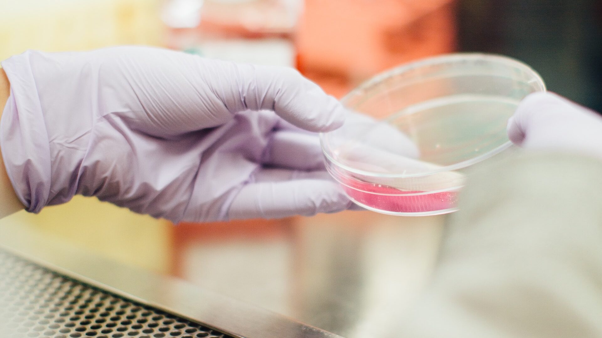
A new study has shown that most cancer cells grown in vitro have little in common genetically with cancer cells in humans.
Human cancer cells grown in culture dishes have the least genetic similarity to their human sources, according to a new computer-based technique developed by researchers at John Hopkins.
According to the researchers, the finding should help shift more resources to cancer research models such as genetically engineered mice and balls of human tissue known as ‘tumouroids’ to better evaluate human cancer biology and treatments, and the genetic errors responsible for cancer growth and progress.
“It may not be a surprise to scientists that cancer cell lines are genetically inferior to other models, but we were surprised that genetically engineered mice and tumouroids performed so very well by comparison,” says Patrick Cahan, PhD, associate professor of biomedical engineering at The Johns Hopkins University and the Johns Hopkins University School of Medicine and lead investigator of the new study.
The new computer modelling technique, CancerCellNet, compares the RNA sequences of a research model with data from a cancer genome atlas to see how closely the two sets match up.
On average, genetically engineered mice and tumouroids have RNA sequences most closely aligned with the genome atlas baseline data in 4 out of every 5 tumour types they tested, including breast, lung and ovarian cancers.
This adds to evidence that cancer cell lines grown in the laboratory have less parity with their human source due to the many differences between a human cell’s natural environment and a laboratory growth environment, the researchers said. “Once you take tumours out of their natural environment, cell lines start to change,” said Prof Cahan.
Around the world, scientists depend on a range of research models to enhance their understanding of cancer and other disease biology, and to develop treatments for conditions. Of these, one of the most widely used is cell lines created by extracting cells from human tumours and growing them with various nutrients in laboratory flasks.
Other methods involve mice that have been genetically engineered to develop cancer, or implanting human tumours into mice, known as xenografting, or use tumouroids.
To investigate the accuracy of these models, scientists often transplant lab-cultured cells or cells from tumouroids or xenografts into mice and see if the cells behave as they should — that is, grow and spread, retaining the genetic hallmarks of cancer. However, the researchers contend that this process is expensive, time-consuming and scientifically challenging and so they developed a more streamlined method. The new technique is based on genetic information about cellular RNA.
“RNA is a pretty good surrogate for cell type and cell identity, which are key to determining whether lab-developed cells resemble their human counterparts,” said Prof Cahan. “RNA expression data is very standardised and available to researchers, and less subject to technical variation that can confound a study’s results.”
To start, Prof Cahan and his team had to choose a standard set of data that acted as a baseline to compare the research models. They used data from The Cancer Genome Atlas as ‘training’ data, which includes RNA expression information of hundreds of patient tumour samples, and other information on the tumour.
They also tested their CancerCellNet tool by applying it to data where the tumour type was already known, such as from the International Human Genome Sequencing Consortium.
The John Hopkins researchers combed through The Cancer Genome Atlas data to select 22 types of tumours for study, and used that data as the baseline for comparing RNA expression data from cancer cell lines, xenografts, genetically engineered mouse models and tumouroids.
Some differences observed included prostate cancer cells from a line called PC3 that started to look genetically more like bladder cancer, Prof Cahan noted. It’s also possible, he said, that originally the cell line was simply labelled incorrectly, or else it could have in fact been derived from bladder cancer. But, from a genetic standpoint, the prostate cancer cell line was not a representative surrogate for what happens in a typical human with prostate cancer.
According to a 0-1 scoring method, cell lines had, on average, lower scoring alignment to atlas data than tumouroids and xenografts.
Prof Cahan said he and his team will be improving the reliability of CancerCellNet by adding additional RNA sequencing data.
Source: John Hopkins Medicine
Journal information: Da Peng et al, Evaluating the transcriptional fidelity of cancer models, Genome Medicine (2021). DOI: 10.1186/s13073-021-00888-w

