Day: May 20, 2021

SA Medical Insurance Schemes in the Crosshairs

The Health Professions Council (HPCSA) said that South Africa’s new National Health Insurance (NHI) should be the sole funding mechanism for health in South Africa.

Addressing parliament on Tuesday, the president of HPCSA, Professor Simon Nemutandani, said that while the organisation accepts that the existence of private medical aid schemes in South Africa can continue, they should funded separately — over and above tax paid for the NHI.

The NHI itself should be funded through taxes paid by all employed South Africans, he said.

“For the NHI to succeed, health must be an exclusive national competence – and any sections of the Constitution that militate against this view must be amended,” the HPCSA stated.

“The Medical Schemes Act must also be amended to ensure alignment with the NHI. NHI should be about funding and contracting, while service provision is left to other entities — public and private.”

Prof Nemutandani said that the NHI Bill should repeal the Medical Schemes Act in its entirety, as the nationalised, centralised health funding system would have no place for it.

For those seeking additional insurance for health cover, they could apply for it under the Insurance Act. Medical schemes should also offer only complementary coverage for services that would not be covered by the NHI, he said.

Additionally, the current reserves of medical schemes — some R90 billion — and all other assets under their control should be transferred to the NHI, said Prof Nemutandani.

“It should be clear that the (NHI) replaces all funding mechanisms for health,” Prof Nemutandani said. “It must also be clear that the NHI is taking over from the medical schemes, and that all assets under the control of the medical schemes must be taken by over the NHI.”

Problematic aspects

The Board of Health Care Funders (BHF) said in its submission that current medical cover providers should be allowed to continue as insurance products. They also pointed out that a number of the Bill’s aspects are problematic, including a provision in the Bill transferring powers and duties of provinces to national government.

The BHF also expected there would be challenges from healthcare service providers and from members of the public over restrictions of their choices. Duplication of services and waste was another concern.

The NHI Bill was presented to and approved by cabinet in July 2019, and has been presented to parliament’s health portfolio committee.

Since then, it has been through an extensive public consultation process through committee roadshows and is scheduled for further parliamentary debates before being presented to the president for promulgation.

However, the Council for Medical Schemes has acknowledged that South Africa’s current financial situation and the impact of the COVID lockdown will make the rollout of the new NHI more difficult.

Source: BusinessTech

More information: Summary of all submissions (PDF)

Neural Connectivity can Predict Epilepsy Outcomes

Image source: Unsplash

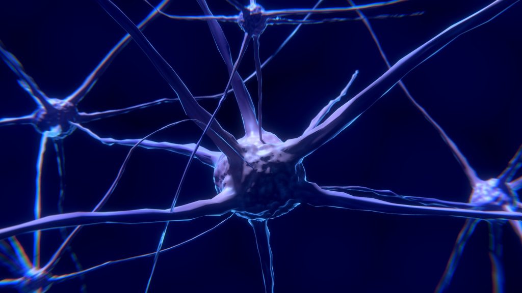
Researchers have found that neuron connectivity patterns within brain regions can better indicate disease progression and treatment outcomes for people with brain disorders such as epilepsy.

Many brain diseases lead to cell death and the removal of connections within the brain. A team led by Dr Marcus Kaiser from the School of Medicine at the University of Nottingham looked at epilepsy patients undergoing surgery. Their findings were published in Human Brain Mapping.

They found that changes in the local network within brain regions can predict disease progression, and also whether surgery will be successful or not.

The team found that looking at connectivity within regions of the brain, showed superior results compared to only observing fibre tract connectivity between brain regions, which is the current method. Dividing the surface of the brain into 50 000 network nodes of comparable size, each brain region could be studied as a local network with 100-500 nodes. There were distinct changes seen in these local networks in patients suffering from epileptic seizures.

Employing diffusion tensor imaging, a special measurement protocol for MRI scanners, the team of scientists showed that fibres within and between brain regions are removed for patients.

However, they found that connectivity within regions better predicted whether surgical removal of brain tissue was successful in preventing future seizures.

Dr Kaiser, Professor of Neuroinformatics at the University of Nottingham, explained: “When someone has an epileptic seizure, it ‘spreads’ through the brain. We found that local network changes occurred for regions along the main spreading pathways for seizures. Importantly, regions far away from the starting point of the seizure, for example in the opposite brain hemisphere, were involved.

“This indicates that the increased brain activity during seizures leads to changes in a wide range of brain regions. Furthermore, the longer patients suffered, the more regions showed local changes and the more severe were these changes.”

The researchers from the involved universities, along with the company Biomax, evaluated the scans of 33 temporal lobe epilepsy patients and 36 control subjects.

Project partners used the NeuroXM™ knowledge management platform to develop a knowledge model for high-resolution connectivity with more than 50 000 cortical nodes and several millions of connections and corresponding automated processing pipelines accessible through Biomax’s neuroimaging product NICARA™.

Project manager Dr Markus Butz-Ostendorf from Biomax said: “Our software can be easily employed at hospitals and can also be combined with other kinds of data from genetics or from other imaging approaches such as PET, CT, or EEG.”

Professor Yanjiang Wang, who is one of the corresponding authors, and Ms Xue Chen, both from China University of Petroleum (East China), commented: “Local connectivity was not only better in overall predictions but particularly successful in identifying patients where surgery did not lead to any improvement, identifying 95% of such cases compared to 90% when used connectivity between regions”.

Source: University of Nottingham

Why Nerves Fail to Regenerate

Image source: Pixabay

Though there are many reasons why nerves fail to regenerate, researchers from Ruhr-Universität Bochum (RUB) have made a breakthrough in their discovery a new mechanism which could lead to effective treatments.

Damage to nerve fibers in the central nervous system – brain, spinal cord, or optic nerve– often results in lifelong and severe disabilities, such as paraplegia or blindness. Though there are various known reasons why nerves fail to regenerate, treating them has not thus far not resulted in success.

Now, the RUB researchers have discovered that nerves release a protein at the injury site that attracts growing nerve fibres — and keeps them entrapped there. This prevents them from growing in the right direction to bridge the injury. Their findings are published in the journal Proceedings of the National Academy of Science (PNAS).

There are three known main causes for the inability of injured nerves of the central nervous system (CNS) to regenerate: insufficient activation of a regeneration program in injured nerve cells that stimulates the growth of fibres, so-called axons; scar formation at the injury site that is difficult for nerve fibres to penetrate; and an inhibitory effect of molecules in the nerve on regrowing axons. “Although experimental approaches have been found in recent decades to address these individual aspects by therapeutic means, even combinatorial approaches have shown only little success,” said Fischer. “So there must be other yet unknown causes for why nerve fibres in the CNS don’t regenerate.”

Using the optic nerve as a model, the research time has now shown another — quite surprising — cause for the regenerative failure in the CNS. The underlying mechanism is not based on inhibition of axon growth, as in the previously identified causes, but instead on a positive effect of a protein at the injury site on the nerve. This molecule is a so-called chemokine known as CXCL12. “The protein actually promotes the growth of axons and attracts regenerating fibers. It is, therefore, chemoattractive,” explained lead investigator Professor Dietmar Fischer. However, this chemoattraction turned out to be more hindrance than help after nerve injury in living animals.

Nerve fibres are trapped

The scientists showed that this protein is released at the nerve’s lesion site and, as a result, keeps the axons at the injured area through the chemoattractive effect. As a result, even some fibres that had already regenerated across the injury site reversed direction, growing backwards to the injury site. The regrowing fibers thus remained trapped due to CXCL12’s attractive effect.

The researchers figured out this effect when they knocked out the receptor for CXCL12 in the retinal nerve cells, rendering them blind to this protein. “Surprisingly, this led to greatly increased fibre growth in the injured optic nerves, and axons showed significantly less regrowth back to the injury site,” Dietmar Fischer points out.

New drug possibilities

The researchers then investigated where at the injury site the CXCL12 originated. They found out that about eight percent of the nerve cells in the retina produce this protein themselves, transport it along their fibers to the injury site in the optic nerve, and release it there from the severed axons. “It is still unknown why some of these nerve cells make CXCL12 and others make the receptor,” said Prof Fischer. “We don’t yet understand the physiological role of the protein, but we can see that it is a major inhibitor of neural repair.”

In further experiments, the researchers showed that knocking out CXCL12 in retinal nerve cells to prevent its release at the injury site equally improved axonal regeneration into the optic nerve. “These new findings open the opportunity to develop pharmacological approaches aimed at disrupting the interaction of CXCL12 and its receptor on the nerve fibres, to free them from their captivity at the site of injury,” concluded Prof Fischer.

His team is now investigating whether similar approaches can also promote the regeneration of axons in other areas of the injured brain or spinal cord.

Source: Ruhr-Universität Bochum

Journal information: Alexander M. Hilla, et al. CXCR4/CXCL12-mediated entrapment of axons at the injury site compromises optic nerve regeneration, in: PNAS, 2021, DOI: 10.1073/pnas.2016409118

Second Gen COVID Vaccine Considered for Booster Shot in SA

Image source: Pexels/CC0

A second generation viral vector COVID vaccine candidate from ImmunityBio Inc is being considered as a booster shot in a study involving nearly 500 000 South African health workers already innoculated with the Johnson & Johnson vaccine.

The health workers, who are the first outside of much smaller studies to receive vaccinations in South Africa, will need a booster, Glenda Gray, the co-lead of the South African studies, said in an interview Wednesday.

“It could be the universal boost that we are looking for,” she said. “Hopefully we will start in a couple of weeks.”

ImmunityBio’s second generation COVID vaccine candidate uses an hAd5 virus as vector. It induces both short term and long term immunity, and besides targeting the coronavirus’ spike proteins like first generation vaccines, it also targets the nucleocapsid protein, which has a lower rate of mutations. Additionally, this hAd5 vector virus provokes an anti-SARS-CoV-2 response, even in individuals with adenovirus immunity.

The magnitude of this T cell response was equivalent to those seen for spike and nucleocapsid T cell responses from previously infected convalescent SARS-CoV-2 patients.

The robust T cell response to both proteins could make it more effective against strains such as the B.1.351 ‘South African’ variant , ImmunityBio said in a statement earlier this year.

The vaccine is also being assessed to determine the safety and effectiveness of oral, sublingual and subcutaneous administration routes.

ImmunityBio’s vaccine is currently in phase I trials in Cape Town, and the company has signed an agreement with South Africa’s BioVac Institute to produce the inoculation in the country should it win approval.

Source: BusinessTech

SARS-CoV-2 Does Not Alter Human DNA

Image source: Pixabay

Despite controversial claims, the SARS-CoV-2 virus likely does not integrate its genetic material into the genes of humans, according to a study published in the Journal of Virology.

A prior study reported the virus’s genetic material was found to have integrated into human DNA in cells in petri dishes, though the scientists conducting the newer research now say this was resulted from genetic artefacts in the testing.

Study co-lead author Majid Kazemian, a Purdue University assistant professor of biochemistry and computer science, said that this finding has two important implications.

“Relatively little is known about why some individuals persistently test positive for the virus even long after clearing the infection,” he explained. “This is important because it’s not clear whether such individuals have been re-infected or whether they continue to be infectious to others. So-called ‘human genome invasion’ by SARS-CoV-2 has been suggested as an explanation for this observation, but our data do not support this case.

“If the virus was able to integrate its genetic material into the human genome, that could have meant that any other mRNA could do the same. But because we have shown that this is not supported by current data, this should allay any concerns about the safety of mRNA vaccines,” he concluded.

It is indeed possible for virus genetic material to be incorporated into the DNA of humans and other animals, which are referred to as ‘chimeric events’. These have happened over many millions of years; human DNA contains approximately 100 000 pieces of ‘fossil DNA’ from viruses that have been accumulated throughout our evolution, accounting for nearly 10% of the genetic material in our cells. Some viral fragments even play a role in diseases such as cancer.

Recent scientific journal articles have raised controversy by claiming that the SARS-CoV-2 virus can also cause these chimeric events. Even before this study demonstrated it was not the case, the researchers suspected it was unlikely, said co-lead author Dr Ben Afzali, an Earl Stadtman Investigator of the National Institutes of Health’s National Institute of Diabetes and Digestive and Kidney Diseases.

“While an earlier study suggested that, in cells infected with SARS-CoV-2, genetic material from the virus copied and pasted itself into human DNA, our group thought this seemed unlikely,” Dr Afzali said. “SARS-CoV-2, like HIV, has its genetic material in the form of RNA but, unlike HIV, does not have the machinery to convert the RNA into DNA. SARS-CoV-2 is unlikely to paste itself into the genome and coronaviruses, in general, does not go near human DNA. As our study shows, we find it highly improbable that SARS-CoV-2 could integrate into the human genome.”

Christiane Wobus, associate professor of microbiology and immunology at the University of Michigan Medical School, another co-lead author on the study, said that the collective understanding of RNA viruses is that SARS-CoV-2 integrating into the human genome was extremely unlikely, it was still worth asking the question.

“Unexpected findings in science—when confirmed independently—lead to paradigm shifts and propel fields forward. Therefore, it is good to be open-minded and examine unexpected results carefully, which I believe we did in our study,” she said. “However, we did not find conclusive evidence for SARS-CoV-2 integration, but instead showed that during the RNA sequencing methodology, chimeras are produced at a very low level as an artifact of the laboratory technique.”

To examine any possible integration event, the researchers came up with a novel technique involving extraction the genetic material from infected cells and then boosting the amount of the genetic material 30-fold. With any chimeric events in the host cell DNA, these bits of genetic material from SARS-CoV-2 would have also increased those by 30. The data did not show this.

“We found the frequency of host-virus chimeric events was, in fact, not greater than background noise,” Kazemian stated. “When we enriched the SARS-CoV-2 sequences from the bulk RNA of infected cells, we found that the chimeric events are, in all likelihood, artifacts. Our work does not support the claim that SARS-CoV-2 fuses or integrates into human genomes.”

Source: Medical Xpress

Journal information: Bingyu Yan et al, Host-virus chimeric events in SARS-CoV2 infected cells are infrequent and artifactual, Journal of Virology (2021). DOI: 10.1128/JVI.00294-21

Inflammation a Predictor of Future Depression in Widowed Spouses

Researchers at Rice University have found that future depression in widowed spouses can be predicted by bodily inflammation after the death of their partners.

The study will be published in the June 2021 edition of the journal Psychoneuroendocrinology. The study was led by lead author Lydia Wu, a Rice psychology graduate student, and Christopher Fagundes, associate professor of psychology and principal investigator for the Biobehavioral Mechanisms Explaining Disparities (BMED) lab at Rice. The researchers recruited 99 participants who had lost their spouses within 2-3 months of the study, and evaluated them on a number of factors, including physical and mental health, over three months.

“Prior research has already linked bodily inflammation to a host of health issues, including cancer, memory issues, heart problems and depression,” Wu said. “We were interested in how systemic inflammation affects the mental health of spouses after losing a loved one. In particular, can inflammation help us identify who will experience clinical levels of depression at a future point in time?”

The researchers found that widowed spouses with higher levels of bodily inflammation immediately after the loss of their partners had more severe symptoms of depression three months later compared to those with lower inflammation levels. This was even more pronounced if they didn’t experience significant depression initially.

Prof Fagundes said that it is normal to experience depression following the death of a spouse, and research shows that undergoing psychotherapy right after the event can actually interfere with people’s natural coping ability.

“We know that most people are remarkably resilient,” he said.

In the case of persistent depression, or depression occurring six or more months after a spouse’s death, it may be a sign that clinical intervention is needed, Prof Fagundes said.

“Until this study, it was difficult to know who was at risk for these persistently high levels of depression and grief until the six-month mark,” he said. “This study identifies a potential biomarker that could help us predict who is at greatest risk for long-term repercussions of loss.”

“This information makes early intervention possible,” Wu said. “We can identify at-risk bereaved persons and introduce them to interventions early on to improve their mental health.”

The researchers said more research is needed to determine who might be at greatest risk.

Source: Rice University

Journal information: E. Lydia Wu et al, Inflammation and future depressive symptoms among recently bereaved spouses, Psychoneuroendocrinology (2021). DOI: 10.1016/j.psyneuen.2021.105206