Cardiovascular risk factors are linked to higher risk of heart disease, but it has been unknown how they are related to abrupt closure of the coronary arteries leading to heart attack and sudden cardiac death. A new study by Mass General Brigham Heart and Vascular Institute investigators finds that people with higher modifiable risk factors burden such as high blood pressure, high cholesterol, diabetes, smoking and obesity have more widespread buildup of plaques in major coronary arteries. Furthermore, these modifiable factors were associated with more unstable, high-risk plaques that are prone to rupture and cause heart attacks. Results are published in JACC: Advances.
“The higher the number of vulnerable, or rupture-prone plaques, the greater the chance that one of them will trigger an adverse event,” said senior author Ik-Kyung Jang, MD, PhD, at the Mass General Brigham Heart and Vascular Institute. “Our findings highlight the importance of early, intensive and sustained interventions to control the modifiable risk factors and prevent future events.”
To better understand how modifiable risk factors and non-modifiable risk factors (including age, sex and family history of heart disease) influence dangerous plaque buildup, the research team examined plaque burden and plaque characteristics across all three major coronary arteries. They analysed 534 plaques in 131 patients who underwent optical coherence tomography (OCT), an intravascular imaging modality that can visualise plaques at high resolution.
The researchers reported that patients with more risk factors had more plaques across all three coronary arteries. In addition, greater number of modifiable risk factors was associated with fewer stable plaques and more prevalent vulnerable plaque features such as lipid plaques with thin fibrous caps that are susceptible to disruption. In contrast, non-modifiable risk factors were associated with more stable plaque phenotypes.
The authors note that prospective research involving larger cohorts, including patients who have previously been treated for blocked coronary arteries, is needed to generalise their findings.
As South Africans face rising inflation, pressure from the burden of chronic disease and financial strain, Momentum Health is advocating for a stronger national focus on the prevention dividend: the long-term value created when South Africans use preventative healthcare, wellness programmes, screenings, health assessments and digital tools to manage health risks before they become serious and costly.
This comes as households, employers and the healthcare system face increasing pressure from affordability constraints, chronic disease and mental health challenges. Research referenced by Stellenbosch Business School has estimated that health-related productivity losses cost South Africa around R161 billion a year, driven by absenteeism, presenteeism and untreated mental health challenges.
Momentum Health believes the conversation needs to move beyond healthcare as an expense that pays only when people are ill. Instead, medical aid should be seen as an investment in helping people stay healthier for longer.
“Rather than paying claims after someone becomes ill, modern healthcare needs to focus on helping people understand their risks earlier, take action sooner and build healthier habits that can improve quality of life over time,” says Damian McHugh, Chief Marketing Officer at Momentum Health. “Prevention is one of the most practical ways to support both better health outcomes and greater financial resilience for South Africans.”
Prevention is becoming more urgent
South Africa’s health and economic realities make prevention increasingly important. PwC’s South Africa Economic Outlook notes that South Africa’s real GDP per capita declined by a cumulative 4.2% over the past decade, a signal often associated with pressure on living standards and economic stability.
At the same time, the country continues to carry a significant disease burden. Government and World Health Organisation reporting show that South Africa has made progress in controlling tuberculosis, with TB cases declining significantly since 2015 and treatment coverage improving to 79% in 2023. However, more than half of TB-affected households still face catastrophic costs, showing how illness can deepen financial vulnerability when people are diagnosed late or struggle to access care.
Mental health is also placing pressure on individuals, employers and families. SADAG’s Working Life Survey found that 52% of surveyed employees had been diagnosed with a mental health condition, with depression, stress, anxiety and burnout among the commonly reported conditions.
These figures show why prevention must be personal, practical and accessible,” adds McHugh. “Every member’s health risks are different. The more people know about their own health, the easier it becomes to make informed choices, use the right benefits and take action before problems escalate.”
Healthy habits can create long-term value
Regular health checks, screenings, physical activity, digital healthcare tools and ongoing health management can help members detect risks earlier and make healthier decisions more consistently.
Preventative care can also support employers and the wider economy. When people are healthier, they are more likely to be present, productive and able to participate fully at work and in their communities. This matters in a country where affordability pressures mean many people are looking for healthcare solutions that deliver tangible value, not only financial protection after illness occurs.
Behavioural incentives can play an important role in this shift. When healthy choices are recognised and rewarded, members are more likely to build routines that support their physical, mental and financial wellbeing. This is the principle behind Momentum’s broader health and wellness approach- making it easier for members to take control of their health before problems arise.
Prevention is personal
The next phase of healthcare will be increasingly personalised. Data, health assessments and digital tools can help members better understand their individual health risks and choose the most relevant actions, whether that means completing a screening, increasing physical activity, accessing care digitally or managing a chronic condition more proactively.
“Prevention works best when it is simple, personalised and part of everyday life,” says McHugh. “Our role is to help members connect the dots between their daily habits, their health outcomes and their financial wellbeing. That is how healthcare becomes more than a monthly contribution. It becomes an investment in a healthier future.”
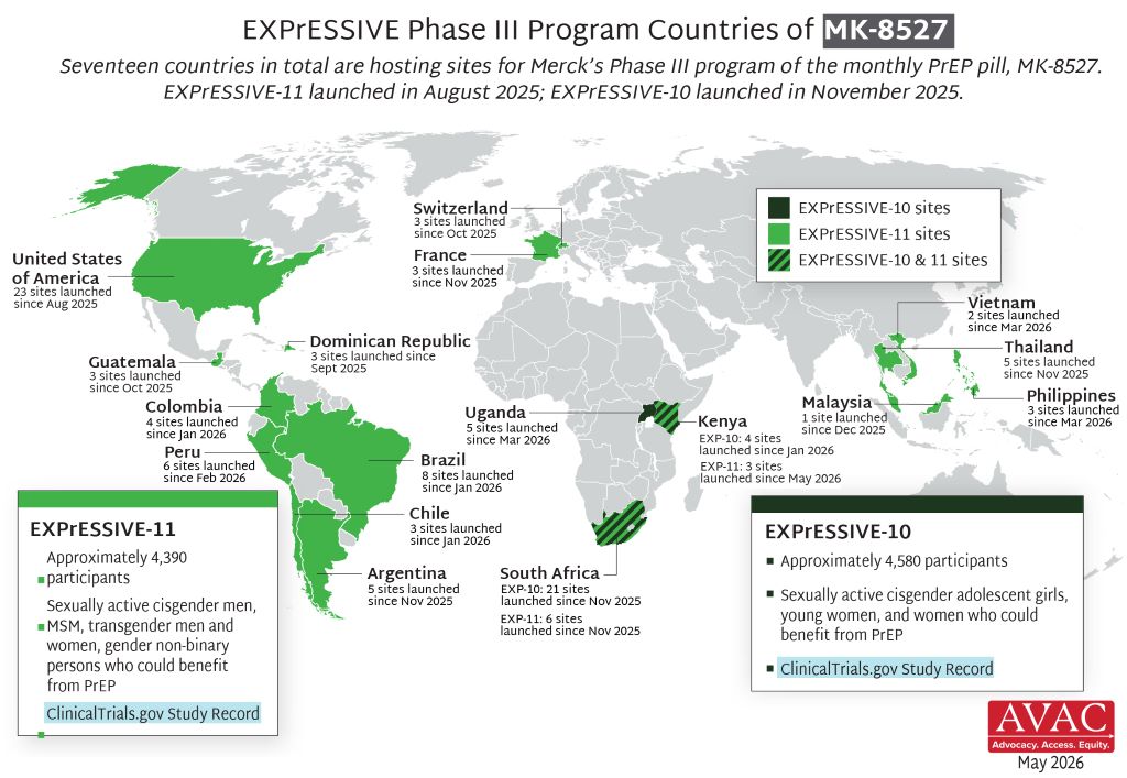
If an HIV prevention pill that provides a month of protection at a time performs well in two ongoing clinical trials, it could become the next big thing in HIV prevention after the lenacapavir injection. A new licensing agreement is paving the way for South Africa’s Aspen Pharmacare to produce the pill should the study findings be positive and the drug be registered.
In June, South Africa’s health department started rolling out the six-monthly lenacapavir HIV prevention injection to around 10% of public sector clinics. While the rollout of this jab still has a long way to go, the next generation of HIV prevention products is already on the horizon.
Two of those new products stand out. One is a new formulation of lenacapavir that looks as if it can provide 12 months of protection at a time. While results so far are promising, the pivotal data on this once-yearly HIV prevention jab is only expected in a year or two.
The other product that has many people in the HIV world excited is a monthly HIV prevention pill that contains a highly potent antiretroviral medicine called alimatravir (it was previously called MK-8527). It starts working within around an hour after someone takes it and appears to provide a month of protection at a time. One benefit of the pill, compared to the lenacapavir injection, is that it would be easier to distribute at scale, given that there is no need for a nurse to administer an injection.
As Spotlight reported in some depth last year, alimatravir looked very promising in a phase 2 study, although for now the jury is still out on the drug’s safety and efficacy. It is currently being evaluated in two pivotal phase 3 clinical trials called EXPRESSIVE-10 and EXPRESSIVE-11. Both these studies started last year and are expected to be completed by around mid-to-late 2027. Medicines are typically only registered for use after positive results in such phase 3 studies.
“The monthly pill offers an alternative for people who would like a long acting, less frequently dosed PrEP but not needle friendly … so it really is about giving more options especially on the pill side,” Professor Linda-Gail Bekker, primary investigator in South Africa on the EXPRESSIVE-10 study, told Spotlight. “You could imagine that just having to remember to take a small easy to swallow pill on the day you pay your bills monthly could be very easy for people. We understand that the packaging is also going to be very user friendly- looking more like a gum packet than a bottle of antiretroviral pills which may also reduce stigma.” (PrEP refers to pre-exposure prophylaxis like HIV prevention pills or injections.)
Licence to make generics
The prospects for future access to alimatravir got a major boost last week when the pharmaceutical company Merck (known as MSD outside of the United States and Canada) announced that it granted licences to seven different companies to produce generic versions of the monthly pill. One of the seven companies is South Africa’s Aspen Pharmacare. The others are Uganda’s Quality Chemical Industries Limited, Kenya’s Universal Corporation Ltd, and Aurobindo, Cipla, Emcure and Viatris in India.
Early responses to the licences have mostly been positive.
Having a generic company with a licence in South Africa is excellent news, said Bekker.
“It is particularly exciting to see manufacturers in Kenya, South Africa and Uganda included in these licenses,” Mitchell Warren, Executive Director of AVAC (a global HIV advocacy group), told Spotlight by e-mail. “These are the first generic PrEP licenses in East and Southern Africa, meaning manufacturing can happen where trials are happening, where need is greatest and where we have the largest PrEP markets.”
“Through our agreement with MSD (Merck), we have the opportunity to support the future supply of an innovative HIV prevention option while strengthening local pharmaceutical manufacturing and healthcare resilience across the continent,” Stephen Saad, Aspen Group Chief Executive, said in a media statement. Under the agreement, the company says it will receive a technical package from Merck, together with licensing rights covering 129 countries, including all African countries.
Speaking to Spotlight, Stavros Nicolaou, Aspen’s Head of Strategic Trade, described alimatravir as “ground-breaking and a potential game-changer”. He commended Merk for starting the licensing process so early. He framed the licence as an important step forward for both South Africa’s HIV response and for local production of antiretrovirals, although he also raised concerns about the procurement of locally manufactured antiretrovirals – the percentage of South Africa’s antiretroviral tenders awarded to local manufacturers has been trending downward.
According to earlier reporting by Business Day, Nicolaou has declined to give any indication as to a potential price for the pill, but he did tell the publication that they could potentially supply it for both South Africa’s public and private sectors.
There are indications that a relatively low price is on the cards. Research being presented at AIDS 2026 this week found that alimatravir could be mass produced and sold at a profit for as little as $15 (around R250 to R300) per person per year. This is less than half the $40 per person per year that South Africa is expected to pay for generic lenacapavir injections in a year or two from now.
“Merck expects to provide initial supply and continue supplying product as needed while licensed generic manufacturers complete development, obtain the necessary regulatory approvals and prepare to provide supply in the licensed territories. The goal is to help avoid delays in access by providing an initial supply pathway until generic manufacturing capacity is established and brought online,” the company said in a media statement.
Earlier licensing
The timing of the licensing announcement is somewhat unusual – such announcements are typically only made after phase 3 trials have been concluded and it is confirmed that the drug is safe and effective.
“Granting licensing agreements to generic manufacturers while clinical trials are still enrolling, before it is known if the product is effective, should significantly reduce the time to market for the product,” Warren said in an earlier AVAC media statement. “The timeline announced today gives us ample opportunity to work with ministries of health, donors, communities, and Merck to plan for broad access to the monthly PrEP pill.”
Warren told Spotlight that the small amount of active drug in alimatravir and the fact that it is an oral dose should make the technology transfer from Merck to generics quite quick. “The hope would be that genetic alimatravir reaches the market within months of the approval of the originator, compared to more than a year for lenacapavir,” he said.
Nicolaou was also upbeat about how quickly things are unfolding. He said that Merck’s decision to execute licences while the phase 3 clinical trials are ongoing allows for an earlier registration pathway (if phase 3 findings are positive, alimatravir will have to be filed for registration with regulators like the South African Health Products Regulatory Authority). He also pointed out that it is a small tablet and that it should be easier to manufacture than HIV prevention injections.
Nicolaou told Spotlight that the plan is for Aspen to do formulation of alimatravir in South Africa, but that they are not currently planning to produce the active pharmaceutical ingredient – this will likely be sourced from Chinese or Indian suppliers.
Some activist criticism
But while the timing has generally been welcomed, there has also been some criticism over the licenses.
A statement from activist group HealthGap points out that Latin American countries like Brazil, Argentina, and Colombia are not included in the list of 129 countries covered by the license, even though some of the phase 3 trial sites for alimatravir are in these countries. The HealthGap statement calls for compulsory licenses to be issued.
In an earlier statement, Merck said that, in recognition of the significant unmet need in Latin America, “Merck is in active discussions with organizations, including Fiocruz (a key player in medicines production and procurement in Brazil), with a goal to enable rapid availability and broad supply of alimatravir in the region”.
Disclosure: The Gates Foundation has provided financial support for clinical trials of alimatravir. Spotlight receives funding from the Gates Foundation, but is editorially independent – an independence the editors guard jealously. Spotlight is a member of the South African Press Council.
An international research team led by Prof. Markus Ege of LMU University Hospital has, for the first time, identified which bacteria in barn air are responsible for the so-called “farm effect,” which can protect against allergies, asthma, and hay fever. Photo by Christopher Stites on Unsplash
An international research team has identified for the first time which bacteria in barn air are responsible for the ‘farm effect’ that can protect against allergies, asthma, and hay fever.
Children who grow up in a farm environment are less prone to allergies, asthma, and hay fever than their classmates. This so-called farm effect has been identified in several observational studies worldwide. It is most likely attributable to the fact that various bacteria present in barn air prevent excessive inflammatory responses of the immune system that are characteristic of such diseases. But which bacteria exactly?
Now, an international team led by Professor Markus Ege from the Dr. von Hauner Children’s Hospital at LMU University Hospital and the Institute of Asthma and Allergy Prevention at Helmholtz Munich has answered this question. The researchers have shown for the first time which specific bacteria in barn air trigger the farm effect, which substances within those bacteria mediate the protection, and which receptors in the body they bind to. Their findings have been published in The New England Journal of Medicine – Evidence.
For years we have been hearing about the hygiene hypothesis, which was first proposed in 1989. This came after three decades of dramatic increases in allergies, asthma, and hay fever among children in Western industrialised countries. These are all conditions in which the immune system mounts an exaggerated inflammatory response and mistakenly attacks the body’s own tissues. According to the hygiene hypothesis, this happens because of under-stimulation in early childhood. The immune systems of children who encounter too few environmental microbes and common cold viruses are more likely to malfunction. “Girls and boys who grow up on farms and are exposed to a wider variety of microbes have the problem far less often,” says Ege. As their immune systems constantly contend with bacterial ‘sparring partners’ from barn air, they are trained to avoid excessive inflammatory responses.
However, the hygiene hypothesis has not been definitively proven, as it is largely based on observational studies. “This kind of research can only show more or less convincing correlations,” says epidemiologist Ege. “But with our new study, we can make a much stronger case, because we can identify the individual links in the proposed causal chain: the bacteria, the relevant microbial metabolic products, and the human receptors.”
The Approach
The researchers analysed data from more than 1000 children participating in European studies in rural areas. For all the children, girls and boys, two types of samples had been collected: nasal swabs and mattress dust. In addition, dust samples were taken from cowsheds for 47 farm children. The team examined which bacteria and fungi were present in these samples, how the different microorganisms were related, and whether specific microbial groups protected the children against asthma.
To this end, the epidemiologists and bioinformaticians employed state-of-the-art genetic and metabolic analyses and developed computer models. Through a process of elimination, they eventually identified the key microorganisms. They also investigated whether the children’s own genes influenced this protective effect. To validate their findings, the researchers additionally used data from France and Finland.
The Results
We identified a small number of bacteria, which we can now pinpoint down to the species level,” explains Giulia Pagani, first author of the study and bioinformatician at the Institute of Asthma and Allergy Prevention at Helmholtz Munich, “such as Romboutsia timonensis and Glutamicibacter arilaitensis. These gram-positive bacteria together mediate two-thirds of the entire farm effect for asthma protection and half of the effect for hay fever and atopic eczema.” The bacteria originate in the cow’s digestive tract, where they produce or metabolize substances like kynurenine, xanthine, alpha-linolenic acid, and stearidonic acid. These compounds are easily inhaled and recognized by two receptors on human airway cells – AhR and PPARγ. “These receptors,” Ege continues, “have multiple functions, including in the immune system, where they appear to prevent excessive inflammatory responses.” Their role in the farm effect was previously unknown. For the first time, therefore, the complete biological chain is visible: cow → barn air → bacteria → metabolic products → human receptors → protection against asthma.
The new findings can now be used by laboratory researchers to decipher the molecular and cellular mechanisms of the farm effect. This opens up the prospect of developing a drug that mimics the farm effect – without the need for children to spend time in barns.
Original publication
G. Pagani et al.: Gram-Positive Bacteria and the Inverse Association between Farm Exposure and Childhood Asthma, NEJM Evidence
Researchers from the Wits Vaccines and Infectious Diseases Analytics (Wits VIDA) Research Unit at the University of the Witwatersrand (Wits) have played a leading role in a landmark international phase 1 / 2 clinical trial demonstrating the safety and immunogenicity of an investigational maternal vaccine against Group B Streptococcus (GBS) – a major cause of life-threatening infections in newborn babies worldwide. The findings have been published in Nature Medicine.
The study evaluated Pfizer’s investigational hexavalent Group B Streptococcus conjugate vaccine candidate (GBS6) in both non-pregnant women and pregnant women, together with their infants. The trial found that the vaccine was generally well tolerated, generated robust immune responses against all six clinically important GBS serotypes, and resulted in efficient transfer of GBS-specific antibodies from vaccinated mothers to their babies.
Wits VIDA was central to the implementation and successful conduct of the clinical trial. South Africa was the primary recruiting country, contributing the overwhelming majority of study participants across both the early and later stages of the trial, reflecting the country’s internationally recognised expertise in maternal and infant vaccine research.
Group B Streptococcus is one of the leading causes of neonatal sepsis, meningitis and pneumonia, accounting for approximately 300 000 serious infections, including 100 000 deaths, in young infants globally each year. The greatest burden of GBS disease in infants occurs in Africa and other low- and middle-income countries, with South Africa reporting among the highest incidence globally (approximately 2-3 cases for every 1000 births). Also, maternal GBS infection could predispose to preterm labour and stillbirths. Current prevention strategies based on intrapartum antibiotics have important limitations, particularly in resource-constrained settings, and do not protect against late-onset disease. Consequently, the WHO has designated a vaccine against GBS, to be given to women during pregnancy aimed at protecting their young infant, as a priority vaccine for development.
Professor Shabir Madhi, Director of Wits VIDA and one of the study’s senior investigators, said: “This publication represents another important milestone in the decades-long effort to develop an effective maternal vaccine against Group B Streptococcus. Maternal immunisation offers the opportunity to protect newborns during the most vulnerable first months of life, when they are at greatest risk of severe infection. These encouraging findings provide further evidence supporting continued development of this vaccine.”
The trial demonstrated comparable safety outcomes between vaccine and placebo recipients. Among pregnant participants, rates of adverse events, serious adverse events and delivery outcomes were similar between the two groups. Infants born to vaccinated mothers likewise had similar safety outcomes to those born to mothers receiving placebo. Importantly, vaccinated mothers developed high concentrations of antibodies that crossed the placenta efficiently, resulting in robust antibody levels in their infants at birth.
The publication builds on Wits VIDA’s longstanding leadership in vaccine research, which has contributed to the development and evaluation of vaccines against respiratory syncytial virus (RSV), rotavirus, pneumococcal disease, influenza virus, COVID-19 and now Group B Streptococcus. The unit continues to play a pivotal role in generating evidence that informs global maternal and child health policy.
The study also highlights the importance of South Africa’s clinical research infrastructure and its contribution to advancing vaccines designed to address diseases that disproportionately affect African populations.
While additional studies are required before the vaccine can be licensed, the results provide strong support for continued clinical development of maternal GBS vaccination as a strategy to reduce newborn deaths and severe infections worldwide.
Wits is currently enrolling in a phase 3 trial for GBS6 (NCT: NCT07160244).
About Wits VIDA
The Vaccines and Infectious Diseases Analytics (VIDA) Research Unit at the University of the Witwatersrand is one of the world’s leading centres for vaccine and infectious disease research. VIDA conducts internationally recognised clinical trials, epidemiological studies and translational research focused on reducing the burden of infectious diseases, particularly among mothers, infants and children in Africa. Its research has contributed to the development and implementation of several life-saving vaccines globally.
By Dr Fatima Hoosain, specialist surgeon and Principal of Apffelstaedt, Hoosain & Associates, with a clinical focus on breast and endocrine surgery.
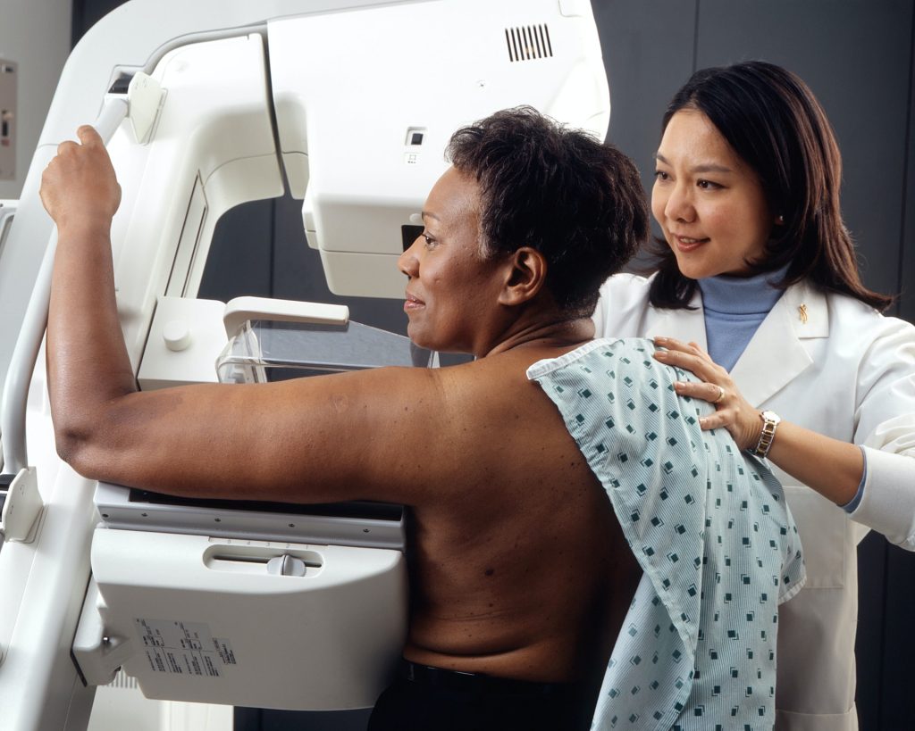
Photo by National Cancer Institute on Unsplash
When people think about breast cancer screening, the conversation often begins and ends with one message: screen more women.
As clinicians, we know it is not quite that simple.
There is no question that screening saves lives. Regular mammography reduces breast cancer mortality and gives us the opportunity to diagnose disease when it is smaller, more treatable and associated with significantly better outcomes. Few interventions in medicine demonstrate such a clear benefit.
The challenge is that good breast care is not defined simply by how many mammograms we perform. It is defined by the quality of the decisions that surround them.
This was the focus of my presentation at the recent Board of Healthcare Funders (BHF) Conference, where we explored how clinicians can balance the burden of breast cancer with evidence-based screening decisions while remaining mindful of both underdiagnosis and overdiagnosis.
Those competing risks are encountered by every clinician involved in breast care.
We all worry about the patient whose cancer is diagnosed later than it should have been. Earlier diagnosis frequently means less extensive surgery, more treatment options and, ultimately, better outcomes. The survival difference between early-stage and advanced disease is substantial, making timely diagnosis one of the most important contributors to long-term prognosis.
At the same time, screening is not without consequences.
Not every abnormality detected on imaging will become life-threatening, yet every suspicious finding understandably creates anxiety. Additional imaging, biopsies and sometimes treatment may follow. Our responsibility is therefore not simply to detect abnormalities, but to interpret them appropriately within the context of each patient’s overall clinical picture.
This is why breast screening should never be approached as a uniform process. Risk matters.
A woman with an inherited genetic mutation or a strong family history should not necessarily follow the same screening pathway as someone at average risk. Likewise, imaging should answer a clinical question. Mammography remains the cornerstone of breast screening, but dense breast tissue, patient age and individual risk factors may require supplementary investigations such as ultrasound or MRI. More imaging is not automatically better medicine. Appropriate imaging is.
These decisions have become even more complex within the South African healthcare environment. International guidelines provide an excellent evidence base, but they do not remove the practical realities we face every day. Access to imaging differs between regions. Advanced investigations may not always be readily available. Medical scheme funding, co-payments and affordability inevitably influence what is possible for many patients. These factors cannot be ignored when discussing best practice because they form part of the reality in which clinical decisions are made.
Fortunately, the treatment landscape continues to evolve.
Advances in oncoplastic surgery, targeted therapies, immunotherapy and modern radiation techniques have transformed outcomes for many patients diagnosed with breast cancer. These developments are encouraging, but they should not distract us from one fundamental principle: the earlier we diagnose clinically significant disease, the greater the opportunity to offer patients treatments that are both effective and less invasive.
Diagnosis, however, is only the beginning of the journey.
Long-term follow-up remains an essential part of breast cancer care. Ongoing surveillance, adherence to endocrine therapy where appropriate, management of treatment side effects and supporting patients through the psychological and financial impact of a cancer diagnosis all influence outcomes. Good breast care extends well beyond the operating theatre or oncology unit.
As our healthcare system continues to face increasing clinical and financial pressures, I believe we need to move beyond simplistic conversations about screening uptake alone.
The more important discussion is whether we are making consistently good clinical decisions. Are we identifying the patients who stand to benefit most? Are we investigating appropriately? Are we avoiding unnecessary intervention when the evidence suggests it is unlikely to improve outcomes?
Those are not easy questions, but they are the ones that matter.
Ultimately, breast cancer screening is not about doing more. It is about doing what is right for the patient sitting in front of us. That remains the most important clinical judgement we make.
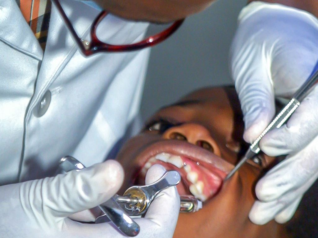
Every year, untreated tooth decay sends thousands of young children to emergency departments for dental problems doctors can’t treat. Many eventually undergo surgery under general anaesthesia, while others endure pain and infection.
A simple, inexpensive liquid called silver diamine fluoride, or SDF, could spare many of those children. Applied to a cavity with a tiny sponge-tipped applicator in about a few second’s time per tooth, SDF arrests decay without drilling, shots or sedation.
Dentists have used SDF successfully for decades in many countries, and off label in the United States since 2014, when it was approved as a medical device to treat tooth sensitivity. However, it has lacked the large US population clinical trials for efficacy and safety that are needed for FDA approval as a drug to treat cavities.
Now, a University of Michigan-led clinical trial has produced that evidence.
Published in JAMA Pediatrics, the Phase III trial enrolled 830 children under age 6 who were recruited through dental offices, pediatric medical practices, Head Start and Early Head Start programs in Michigan, New York and Iowa.
Researchers found that 38% SDF arrested tooth decay in more than half of children’s affected baby teeth when treated at 6-month intervals. Unlike conventional treatment, which removes part of the tooth before placing a filling, SDF is simply painted onto the cavity.
“This is a very effective and safe treatment – even in children as young as 1,” said Margherita Fontana, professor of dentistry at the University of Michigan School of Dentistry and the study’s lead investigator.
Tooth decay is the most common chronic disease of childhood, affecting more than 40% of US children. Left untreated, cavities can cause severe pain, infection, difficulty eating and sleeping, missed school and repeated medical visits.
SDF may be especially valuable for very young children, older adults, people with developmental or physical disabilities, patients with severe dental anxiety, and others who cannot easily tolerate or access conventional dental treatment, Fontana said.
Its primary drawback is cosmetic, she said. The silver permanently darkens the decayed portion of the tooth.
“If we want more children and families to benefit from this treatment, we need rigorous evidence showing both that it works and that it’s safe. From a public health perspective, if we want broader implementation across the United States, including in medical settings, we need carefully collected data in U.S. populations, and we now have that,” Fontana said.
The study began in 2018 and progressed even with the challenges of the COVID-19 pandemic.
“In medicine, clinicians want high-quality evidence before changing practice,” Fontana said. “It is important to have data they can refer to because young children often see paediatricians years before they ever visit a dentist, broader acceptance could allow many more cavities to be treated while a referral to a dental home is successful, and before they become painful, infected or require surgery.”
The product used in this trial, Advantage Arrest 38% SDF, was provided by Elevate Oral Care.
Amr Moursi, professor of paediatric dentistry at New York University College of Dentistry, said the study provides important data for broadening use of SDF.
“Our results support FDA approval of SDF for managing arrest of tooth decay in young children. Removing SDF from off-label status would be an important innovation which could lead to increased utilisation by providers, enhanced payments by insurers and more consistent product quality,” said Moursi, a co-principal investigator on the study.
For some children, reapplying SDF every few months may be all that’s needed until the baby tooth naturally falls out. For adults, it may serve as a long-term treatment or as a bridge until restorative procedure is affordable or practical.
“For almost anyone, this can arrest the decay and stop the infection and the pain it causes,” Fontana said. “This could benefit many people.”
A clinical trial led by researchers from Hospital Clínic de Barcelona, the August Pi i Sunyer Biomedical Research Institute (IDIBAPS) and The Institute for Advanced Chemistry of Catalonia (IQAC-CSIC) has compared the effectiveness of abatacept and hydroxychloroquine in preventing the development of rheumatoid arthritis in patients with palindromic rheumatism, an autoimmune disease that progresses to arthritis in approximately half of all patients. The trial was conducted over two years across 14 hospitals throughout Spain and involved 70 patients with palindromic rheumatism. The findings, published in Nature Medicine, indicate that abatacept is significantly more effective than hydroxychloroquine in preventing the onset of arthritis.
Palindromic rheumatism is characterised by intermittent episodes of joint inflammation, with acute flare-ups lasting a few days and resolving spontaneously. However, around half of patients eventually develop rheumatoid arthritis, a chronic disease that causes irreversible joint damage. This risk is particularly high in individuals with biomarkers such as rheumatoid factor and anti-citrullinated peptide antibodies. The presence of these two autoantibodies – proteins of the immune system that mistakenly attack the body’s own organs and tissues – is used in the diagnosis of the disease.
Until now, in the absence of clinical trial evidence, the standard approach has been to treat patients with palindromic rheumatism using hydroxychloroquine, a drug with anti-inflammatory and immunosuppressive properties aimed at improving disease symptoms. In this context, the team led by Raimon Sanmartí, head of the Inflammatory Arthropathies Research Group at IDIBAPS, conducted a two-year clinical trial involving 70 patients with palindromic rheumatism. The objective was to compare the effectiveness of hydroxychloroquine with abatacept, a lymphocyte inhibitor – a type of white blood cell that attacks the joints after mistakenly identifying them as a threat – in reducing progression from palindromic rheumatism to rheumatoid arthritis
The results show that treatment with abatacept significantly reduces progression to rheumatoid arthritis. Only 20% of patients receiving abatacept developed arthritis, compared with 50% of those treated with hydroxychloroquine. Furthermore, patients treated with abatacept not only avoided progression to rheumatoid arthritis in most cases, but also experienced a significant improvement in palindromic rheumatism symptoms. “The study shows that patients treated with abatacept are more likely to achieve complete remission of attacks associated with acute pain and joint swelling, and that their inflammatory episodes are less severe,” explains Isabel Haro, Head of the Peptide Synthesis and Biomedical Applications Unit at IQAC-CSIC. The research team also highlights that both drugs proved safe and well tolerated throughout the trial.
Early intervention
“The results of this study indicate that we can intervene at an early stage to modify the natural course of the disease and reduce the risk of patients developing more severe and irreversible conditions,” says Sanmartí. “This opens the door to a paradigm shift in the treatment of these patients.”
The study, which involved several national research centres, also analysed the evolution of a number of biomarkers (autoantibodies) developed by the CSIC research group during patient follow-up. “Although no significant differences were observed between abatacept and hydroxychloroquine in terms of autoantibody responses, this work demonstrates the value of immunomodulatory approaches in the early stages of disease, when it is still possible to prevent progression to more severe and chronic forms,” notes Haro.
Rheumatoid arthritis is a disease that significantly affects patients’ quality of life and places a considerable burden on healthcare systems. Preventing its development in a substantial proportion of cases represents an important advance in the management of rheumatic diseases.
A new study published in the American Academy of Family Physicians finds that keeping patients connected to the same practice and physician over time may help reduce avoidable hospital use.
This retrospective cohort study using longitudinal data from 100 450 patients across 48 general practices in and around Amsterdam found that patients registered with their practice for longer than 5 years had 9% to 21% lower odds of urgent hospital admission and 17%-28% lower hospital costs compared to those registered for 0 to 5 years.
Consistently seeing the same general practitioner was associated with 6% to 7% lower hospital costs, but not with fewer urgent admissions. Researchers measured continuity associations with urgent hospital admissions and hospital costs in two ways: duration of the general practitioner-patient relationship and how concentrated a patient’s visits were with one physician.
The researchers concluded, “Our study suggests an association between continuity in general practice and hospital use and costs. Although there is an overwhelming amount of evidence regarding the benefits of continuity of care for both patients and GPs, this study shows that continuity is also associated with fewer urgent admissions and lower hospital costs.”
By Brian Harris, CEO at Turnberry Management Risk Solutions
28 July 2026 – Medical aid remains essential for accessing private healthcare in South Africa, but it does not always cover the full cost of treatment. While regulations such as Prescribed Minimum Benefits (PMBs) ensure that members have access to a defined level of care for certain conditions, medical schemes still apply tariff limits, treatment protocols, co-payments and other funding rules that can leave members exposed to out-of-pocket costs. At the same time, healthcare costs continue to rise, placing additional pressure on how schemes fund treatment. As a result, medical expense shortfalls are becoming increasingly common, making gap cover an essential part of helping clients manage their healthcare costs and protecting them against unexpected expenses.
Protection within limits
PMBs are a clear example of how scheme rules and funding limits influence what medical schemes ultimately pay for. They are designed to ensure that all medical scheme members have access to treatment for a defined list of emergencies, chronic, and life-threatening conditions. However, they do not provide unlimited funding for every treatment option, and there is often a misunderstanding about this.
When it comes to PMBs, cover is still subject to scheme rules, treatment protocols, formularies, and designated service provider requirements. In many cases, cover is also aligned to the level of care that would ordinarily be available in the public healthcare system. This becomes particularly important in areas such as oncology, where newer or more specialised treatments may fall outside what a scheme is required to fund in full.
As a result, members may still face co-payments, sub-limits, or shortfalls that need to be paid for out of pocket, even when the condition itself qualifies as a PMB.
How scheme rules create shortfalls
PMBs are only part of the picture when it comes to medical expense shortfalls. Even where treatment is covered, medical schemes reimburse according to their own tariffs and funding rules, while healthcare providers may charge significantly more. The difference between what the scheme pays and what the provider charges is the member’s medical expense shortfall.
At the same time, medical schemes use co-payments, benefit limits, Designated Service Providers (DSPs) and authorisation requirements to manage rising healthcare costs and keep contributions affordable. If these rules are not followed, or if treatment falls outside the approved funding structure, members may still need to pay part of the cost themselves.
This means that having medical aid does not always guarantee that treatment will be covered in full. Even where treatment is approved, members can still face significant out-of-pocket expenses.
Advice is essential
As funding rules become increasingly complex, advisers need to take on a more proactive role in helping clients understand and navigate the healthcare landscape. Many clients do not fully understand how PMBs, DSPs, funding rules and scheme tariffs affect what their medical aid will ultimately pay. The reality often only becomes clear at the claims stage, when members discover that they are responsible for part of the cost themselves.
Explaining these rules clearly, together with where medical expense shortfalls may arise, is therefore becoming an increasingly important part of the healthcare funding discussion. Regular reviews and clear guidance also help ensure that changes to scheme rules, benefits, and healthcare costs do not leave clients exposed to unnecessary or unexpected out-of-pocket expenses.
Future-proofing healthcare advice
Medical aid and gap cover should not be treated as once-off decisions. Scheme rules, pricing structures and healthcare costs continue to change, which means healthcare advice needs to be reviewed regularly to ensure that cover remains appropriate.
Helping clients understand how their medical aid works, where medical expense shortfalls may arise, and how gap cover can be structured forms an important part of responsible advice. Regular reviews also help ensure that changes to benefits, family circumstances or healthcare needs do not leave clients exposed to unnecessary costs.
As regulation continues to shape how healthcare is funded in South Africa, gap cover is becoming an increasingly important part of managing healthcare costs. Brokers and financial advisers play an essential role in ensuring their clients have medical aid and gap cover that together provide the right level of protection.
Turnberry Management Risk Solutions (Pty) Ltd is an authorised Financial Services Provider (FSP no. 36571). Underwritten by Lombard Insurance Company, an Authorised Financial Services Provider (FSP 1596) and Insurer conducting non-life insurance business.